


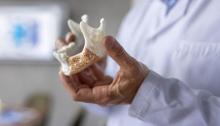
Print the Bone Before You Place the Implant
Customized scaffolds are transforming guided bone regeneration by delivering precontoured, fixation-ready solutions that improve predictability in complex ridge defects.
PURCHASE COURSE
This course was published in the September/October 2026 issue and expires October 2029. The authors have no commercial conflicts of interest to disclose. This 2 credit hour self-study activity is electronically mediated.
This course was published in the September/October 2026 issue and expires October 2029. The authors have no commercial conflicts of interest to disclose. This 2 credit hour self-study activity is electronically mediated.
AGD Subject Code: 690
EDUCATIONAL OBJECTIVES
After reading this course, the participant should be able to:
- Explain the digital workflow and design principles, material selection, and clinical indications of customized three-dimensionally printed scaffolds for implant site development.
- Identify the clinical applications of a customized nonresorbable titanium mesh and customized resorbable bioceramic blocks.
- Discuss the application of the two types of scaffolds.
Alveolar ridge bone volume deficiency is a common barrier to restoratively driven dental implant placement. Conventional bone augmentation techniques, such as guided bone regeneration (GBR), stabilize bone grafts with resorbable or nonresorbable membranes or metallic meshes. However, shaping and fixation are performed intraoperatively and can be time-consuming and less predictable in large vertical or combined defects, particularly for less experienced surgeons.
With the adoption of digital technologies, such as cone-beam computed tomography (CBCT), intraoral scans, and three-dimensional (3D) printing, new solutions emerge that enable clinicians to customize scaffolds to individual patients. Patient-specific 3D-printed scaffolds are designed from CBCT scan data to match defect anatomy and provide a rigid, precontoured framework that maintains space while supporting bone formation.1–5
Restorative-driven dental implant rehabilitation of the full or partial edentulous alveolar ridge requires adequate 3D bone volume to position the implants with stable peri-implant tissues. Following extraction, the alveolar ridge undergoes resorptive remodeling, often greatest on the buccal aspect, with clinically relevant loss of width and height during early healing.6 For many patients, ridge preservation or augmentation becomes the first stage of implant therapy.
While effective, GBR’s predictability in severe defects depends on variables that are difficult to standardize, including intraoperative barrier adaptation, fixation, contouring, and flap management.1,2 In severe vertical or combined defects, small inaccuracies can translate into dead space; graft micromotion, which can lead to complications such as soft-tissue dehiscence; infection; and loss of regenerated bone volume.
With advances in digital dentistry and additive manufacturing, the field of bone regeneration now offers a different approach: define the prosthetic target digitally and fabricate a patient-specific scaffold that delivers the planned contour with planned fixation.
Digital Workflow and Material Selection
For implant site development, a scaffold is a 3D construct placed at a deficient ridge to maintain space, stabilize the graft material, and guide vascular and cellular ingrowth until sufficient native bone forms to support implants.4,7 Patient-specific scaffolds differ from conventional meshes, or bone blocks, because the external geometry and fixation features are designed from the patient’s CBCT dataset rather than adapted chairside.3
CBCT data are exported in DICOM format and segmented to delineate the defect and adjacent roots and neurovascular structures. Prosthetically driven planning (diagnostic wax-up or digital setup) defines the target tooth position and ridge contour, after which the scaffold is modeled to reproduce the planned form while maintaining safety offsets and incorporating preplanned fixation channels. The finalized STL file is fabricated with a material-appropriate additive manufacturing workflow (eg, laser sintering/melting for titanium, fused deposition modeling for thermoplastics, or printing and sintering for calcium-phosphate ceramics).2,4
Material selection determines whether the device behaves primarily as a space-maintaining barrier that is removed (eg, titanium mesh) or as an osteoconductive graft substitute that is remodeled into bone (eg, calcium phosphate or hydroxyapatite blocks).
Customized Nonresorbable Titanium Meshes
Patient-specific titanium meshes are among the most widely used customized devices for ridge augmentation. Titanium provides high strength and stiffness and can be printed to achieve a precise fit and planned contour. As titanium is nonresorbable, the mesh is typically removed at re-entry during implant placement.
Clinical studies report clinically meaningful ridge gains prior to implant placement when customized titanium meshes are combined with particulate grafting and meticulous soft-tissue management.8,9 Design considerations include rounding mesh edges and avoiding over-contouring at anticipated incision lines to reduce exposure risk, incorporating fixation holes to permit rigid stabilization and minimize micromotion, and planning soft-tissue management to achieve tension-free closure. Staged soft-tissue augmentation may be appropriate in patients with thin biotypes.
Customized Nonresorbable Polymer Meshes
Polyether ether ketone (PEEK) is a high-performance polymer that can be fabricated as a patient-specific barrier. It is radiolucent and avoids metallic imaging artifacts, but it is bioinert, nonresorbable, and generally requires removal.9,10 Clinical studies have reported predictable dimensional gains before implant placement with patient-specific PEEK barriers, and a randomized clinical trial comparing customized PEEK vs titanium mesh reported comparable augmentation outcomes when protocols were standardized.10,11
Customized Resorbable Polymer Scaffolds
Resorbable thermoplastics, such as polycaprolactone (PCL), can be printed into patient-specific frameworks. PCL has a slow degradation profile, which can help maintain space during prolonged bone formation, but also extends scaffold presence in the wound.12 In a pilot randomized controlled trial, insertion of a 3D bioresorbable PCL scaffold into extraction sockets improved maintenance of ridge height at 6 months compared with natural healing, with mineralized bone observed within scaffold porosities.12
Patient-specific resorbable scaffolds have also been reported as frameworks for staged augmentation before implant placement, demonstrating regenerated volume adequate for implant insertion and histologic evidence of new bone formation.13 For larger ridge defects, custom PCL mesh frameworks combined with particulate grafting have shown vertical and horizontal gains sufficient to support implant therapy in case reports.14
Customized Resorbable Bioceramic Blocks
Three-dimensionally printed calcium-phosphate ceramics (eg, hydroxyapatite, β-tricalcium phosphate, biphasic calcium phosphate) can be fabricated as patient-specific blocks that function as osteoconductive graft substitutes. These constructs are fixed to native bone as a block and guide bone ingrowth through a porous architecture, with gradual remodeling depending on composition and resorption kinetics.4,15
In a case series of horizontal ridge defects, customized 3D-printed nanohydroxyapatite blocks were designed from CBCT data, fixed to the ridge, and combined with autologous blood-derived concentrates; re-entry at 6 months demonstrated ridge gain, biopsy-based evidence of new bone formation, and successful implant placement.16 Long-term human histology supports that printed calcium-phosphate ceramics can remain integrated and remodeled in the alveolar ridge years after placement.15
In particular, for bioceramic bone blocks, interconnected porosity supports early vascular invasion and diffusion of nutrients, which are prerequisites for robust osteogenesis.5 However, higher porosity reduces mechanical stability.5 Clinically, implant-site scaffolds must balance biologic permeability with stiffness sufficient to resist collapse under soft-tissue pressure, particularly in vertical augmentation.
Clinical Indications and Possible Complications
Customized scaffolds can reduce intraoperative variability by delivering a precontoured framework matched to the planned ridge form. Indications for customized scaffolds include severe horizontal and/or vertical ridge deficiencies requiring precise contour control; defects in which intraoperative mesh bending is difficult (eg, anterior maxilla or combined defects); proximity to anatomical structures, such as mental nerve; and clinical scenarios in which reduced operative time and standardized barrier fit may mitigate technique sensitivity.3
Scaffold exposure remains the predominant complication for space-maintaining barriers and slow-resorbing scaffolds. Risk increases with thin, soft-tissue biotype, limited keratinized tissue, flap tension, over-contouring, smoking, and suboptimal plaque control.2,3 Soft-tissue dehiscence and scaffold exposure can increase contamination and compromise regenerated volume.
Risk mitigation begins at scaffold design with features such as rounded edges, reduced prominence, and continues through surgery with considerations such as rigid fixation, periosteal release for tension-free closure, and meticulous flap handling. In selected cases, autologous blood products (ABP) such as platelet-rich fibrin (PRF) may be used as an adjunct.
Surgical Considerations for Successful Outcomes
Although materials selection and scaffold designs are important for clinical success, clinical considerations at the pre-operative, intra-operative, and post-operative stages determine both the short-term and long-term success of bone augmentation and implant rehabilitation.
Pre-operative planning includes control of systemic inflammation, and local active disease (eg, periodontitis, endodontic infection), along with optimization of plaque control and patient oral hygiene self-care. In addition, prosthetic design to guide implant position and the required ridge dimensions are critical to success. Scaffold should be designed to reproduce the target bone volume and contour with planned fixation and smooth scaffold margins at anticipated flap margins to reduce risks of soft tissue dehiscence. Finally, selection of an appropriate staged timeline based on defect morphology and expected maturation is key.
A full-thickness flap with adequate tension-free release is elevated. The recipient bed is thoroughly degranulated, and cortical perforations (decortication or microperforations) may be performed to enhance bleeding and angiogenesis. The scaffold is secured with fixation screws to minimize micromotion. For barrier-type devices (eg, titanium or PEEK), the contained space is densely filled with particulate graft to balance volume stability and remodeling. For 3D printed ceramic blocks, intimate contact with native bone is critical and particulate graft is limited to fill the residual gaps. Adjunctive soft-tissue protection (eg, collagen membranes, connective tissue grafting, and/or ABP) may be considered.17 Primary, tension-free closure is achieved with periosteal release and layered suturing.
Post-Operative Care and Re-Entry
Post-operative management emphasizes plaque control, antiseptic rinses, and close follow-up during the first 2 to 4 weeks. At re-entry, nonresorbable barriers are removed, regenerated bone is assessed, and implants are placed in a restoration-driven position. Minor contour discrepancies may be corrected with secondary contour bone augmentation. If scaffold exposure occurs, management is determined by timing and severity. Early, extensive exposure with infection often necessitates early removal of the scaffold, whereas late, small exposures without suppuration may be managed with local hygiene measures and careful monitoring until re-entry.
![]() Case Study 1
Case Study 1
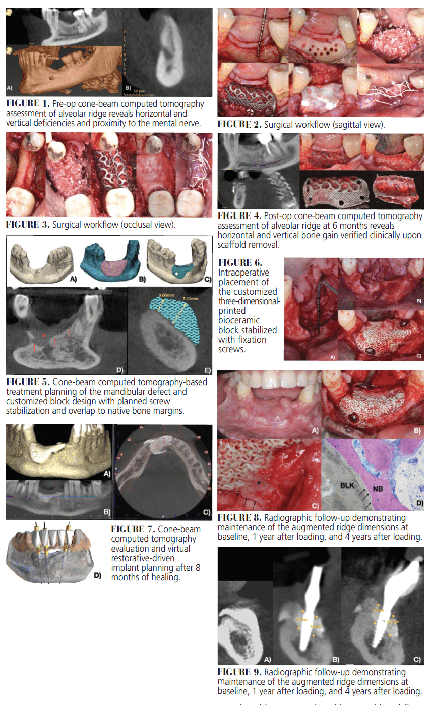
A patient presents with an advanced alveolar ridge defect of the mandible (Figure 1). The patient was treated with GBR utilizing a customized 3D-printed titanium mesh framework in combination with a mixture of allograft and xenograft (Figure 2). The bone graft materials were hydrated with recombinant human platelet-derived growth factor-BB. In addition, the Ti-mesh was covered with a collagen barrier membrane (Figure 3). Backward design was performed to guide and facilitate future implant placement in one of the clinical cases. The CBCT scan at 6 months post-op and clinical assessment upon uncovering of the graft site revealed ridge augmentation and bone formation (Figure 4).
Case Study 2
A patient presented with a severe anterior mandibular ridge defect secondary to gunshot trauma, with > 22 mm vertical deficiency that precluded restoration-driven implant placement. CBCT imaging was used for defect segmentation. A patient-specific porous bioceramic block was digitally designed to restore ridge height and contour and included two screw channels for rigid fixation (Figure 5).
A full-thickness flap was elevated, and the recipient bed was prepared. The customized block demonstrated intimate adaptation to the defect margins and was stabilized with two titanium fixation screws (Figure 6). L-PRF was combined with the construct to support early healing and soft-tissue management. Primary closure was achieved without tension.
At 8 months, CBCT evaluation demonstrated substantial ridge regeneration with maintenance of the planned contour (Figure 7). Digital implant planning was performed, and three implants (3.5 × 13 mm) were placed in the reconstructed ridge. A biopsy harvested during implant site preparation demonstrated osteogenesis adjacent to the bioceramic scaffold, consistent with graft incorporation.
The site was restored with an implant-supported ceramic prosthesis. Follow-up radiographs demonstrated stable peri-implant bone levels and durable maintenance of the augmented ridge dimensions at 18 months and up to 4 years after functional loading (Figures 8 and 9), supporting the feasibility of patient-specific printed bioceramic blocks for severe ridge defects prior to implant rehabilitation.
Discussion
Digital, restoration-driven planning remains fundamental; scaffold design should deliver the target ridge form and incorporate fixation trajectories that respect anatomic constraints. Rigid stabilization is essential to limit micromotion, and soft-tissue management frequently dictates outcomes in vertical or combined defects.
Material selection should be matched to the clinical objective: removable titanium or PEEK meshes optimize space maintenance, whereas patient-specific bioceramic blocks function as osteoconductive graft substitutes in selected defects and may reduce the need for autogenous block harvesting. Current clinical evidence is dominated by case reports and small series; controlled comparative studies are needed to clarify indications, complication profiles, and long-term remodeling across materials.
Conclusion
Patient-specific 3D-printed scaffolds extend GBR by standardizing space maintenance and contour control for implant site development. Customized titanium and PEEK meshes provide strong, predictable frameworks but require removal, while customized bioceramic blocks act as osteoconductive graft substitutes that can integrate and remodel. When paired with prosthetic-driven planning, rigid fixation, and meticulous soft-tissue management, customized scaffolds can support predictable ridge development for restorative-driven implant placement.
References
- Asa’ad F, Pagni G, Pilipchuk SP, et al. 3D-printed scaffolds and biomaterials: review of alveolar bone augmentation and periodontal regeneration applications. Int J Dent. 2016;2016:1-15.
- Rider P, Kačarević ŽP, Alkildani S, et al. Additive manufacturing for guided bone regeneration: a perspective for alveolar ridge augmentation. Int J Mol Sci. 2018;19:3308.
- Elrefaei SA, Parma-Benfenati L, Dabaja R, et al. Customized 3D-printed scaffolds for alveolar ridge augmentation: a scoping review of workflows, technology, and materials. Med (Kaunas). 2025;61:1269.
- Wang C, Huang W, Zhou Y, et al. 3D printing of bone tissue engineering scaffolds. Bioact Mater. 2020;5(1):82-91.
- Karageorgiou V, Kaplan D. Porosity of 3D biomaterial scaffolds and osteogenesis. Biomaterials. 2005;26:5474-5491.
- Araújo MG, Lindhe J. Dimensional ridge alterations following tooth extraction: an experimental study in the dog. J Clin Periodontol. 2005;32:212-218.
- Pilipchuk SP, Plonka AB, Monje A, et al. Tissue engineering for bone regeneration and osseointegration in the oral cavity. Dent Mater. 2015;31:317-338.
- Lee SY, Choi SH, Lee DW. Vertical ridge augmentation with customized titanium mesh using a 3D-printing model: a prospective study in humans. Int J Oral Maxillofac Implants. 2024;39:153-163.
- Tallarico M, Park CJ, Lumbau AI, et al. Customized 3D-printed titanium mesh developed to regenerate a complex bone defect in the aesthetic zone: a case report approached with a fully digital workflow. Materials. 2020;13:3874.
- El Morsy OA, Barakat A, Mekhemer S, Mounir M. Assessment of 3-dimensional bone augmentation of severely atrophied maxillary alveolar ridges using patient-specific poly ether-ether ketone (PEEK) sheets. Clin Implant Dent Relat Res. 2020;22:148-155.
- Mounir M, Shalash M, Mounir S, et al. Assessment of three-dimensional bone augmentation of severely atrophied maxillary alveolar ridges using prebent titanium mesh vs customized polyetheretherketone mesh: a randomized clinical trial. Clin Implant Dent Relat Res. 2019;21:960-967.
- Goh BT, Teh LY, Tan DBP, Zhang Z, Teoh SH. Novel 3D polycaprolactone scaffold for ridge preservation—a pilot randomised controlled clinical trial. Clin Oral Implants Res. 2015;26:271-277.
- Ivanovski S, Staples R, Arora H, et al. Alveolar bone regeneration using a 3D-printed patient-specific resorbable scaffold for dental implant placement: a case report. Clin Oral Implants Res. 2024;35:1655-1668.
- Park JY, Jeon SH, Lee JY, Park JM, Cha JK. Vertical and horizontal ridge augmentation using customized three-dimensionally printed polycaprolactone mesh in atrophic posterior maxillae: a case report. J Oral Implantol. 2025;51:326-336.
- Mangano C, Giuliani A, De Tullio I, et al. Histological and histomorphometrical results of a 3-D printed biphasic calcium phosphate ceramic 7 years after insertion in a human maxillary alveolar ridge. Front Bioeng Biotechnol. 2021;9:614325.
- Mekcha P, Wongpairojpanich J, Thammarakcharoen F, Suwanprateeb J, Buranawat B. Customized 3D printed nanohydroxyapatite bone block grafts for implant sites: a case series. J Prosthodont Res. 2023;67:311-320.
From Decisions in Dentistry. September/October 2026;12(2):32-35.