
Optimal Tissue Positioning With Implants in Smile Makeover Treatment
This case study involves correcting asymmetry with crown lengthening and utilizing implant adaptation with porcelain veneers and zirconia crowns to achieve an esthetic smile makeover.
This case report demonstrates how to achieve optimal esthetic results when placing anterior implants next to porcelain veneers. Ideally, the results are enhanced when there is tissue symmetry, consistent color, and an ideal shape and position of all restorations. The 56-year-old male in this report has been a patient in my practice for nearly 30 years. At an early age, he had endodontic treatment on teeth #9 and 10 (Figure 1). Recently, these teeth needed to be extracted and were replaced with bone grafts and implants (Figure 2).
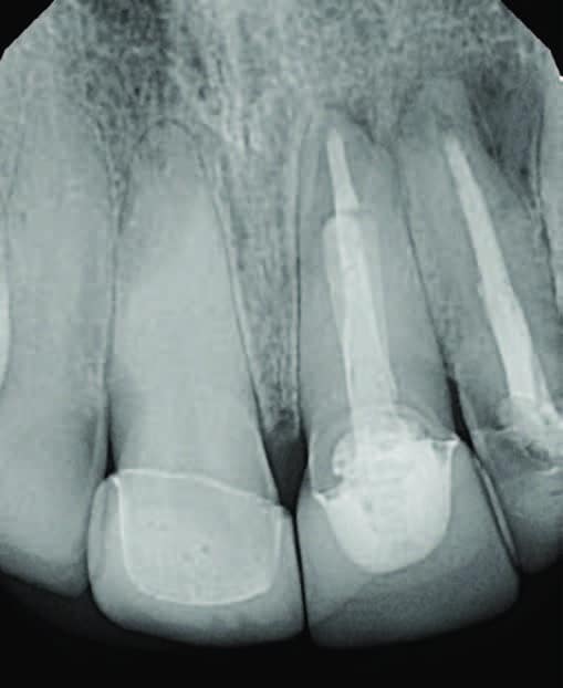
prior to the failure of endodontically
treated teeth #9 and 10.
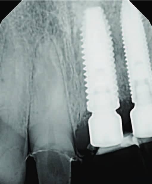
placement of implants at sites #9 and 10.
Once the implants were placed by the periodontist, we waited two months for the tissue to completely heal before starting to restore the case. At the time of implant placement, lab-fabricated custom provisionals were splinted and placed on teeth #9 and 10.
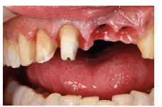
Following a two-month healing process, there is an apparent asymmetry in the patient’s maxillary laterals and centrals (Figure 3).
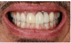
8, 9 and 10.
CROWN LENGTHENING
Tooth #7 had a 20-year-old porcelain veneer and #8 had a full porcelain crown placed at the same time. The patient agreed to return to the periodontist for a crown lengthening procedure on teeth #7 and 8 to establish better symmetry. Furthermore, the final treatment was discussed and the patient agreed to the following:
- Feldspathic porcelain veneers for teeth #6, 7 and 11
- Zirconia crowns on teeth #8, 9 and 10
Crown lengthening was completed and, after two months of healing, new temporary restorations were placed on teeth #7 and 8 (Figure 4).
PREPARATION
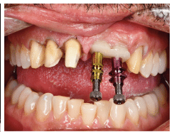
At the preparation appointment, porcelain veneer preps were completed on teeth #6, 7 and 11, and a crown prep was refined on #8. The two-unit temporary was then removed from teeth #9 and 10 (Figure 5).
RESTORATIVE TREATMENT
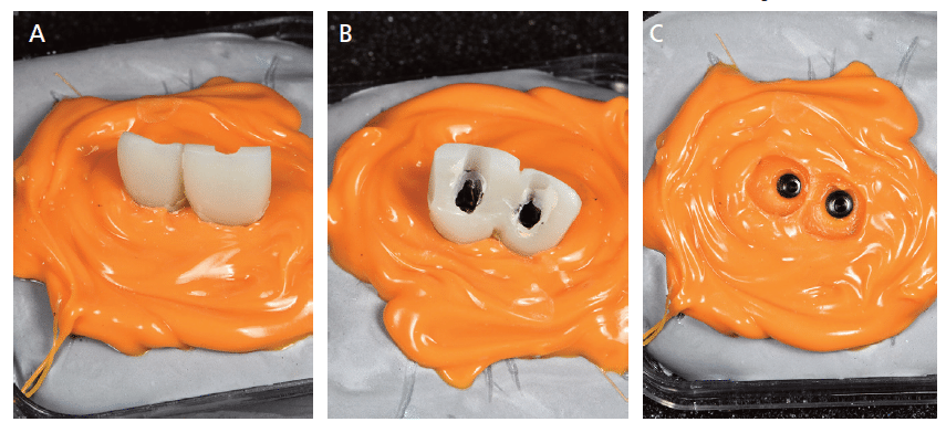
Next, lab analogs were attached to the custom temporaries and secured in stone (Figures 6A and 6B). Once the stone had set, polyvinyl impression material was flowed around the gingival one-third of the temporaries to capture their shape so the lab could produce a precise replica to ensure accurate tissue placement with the new restorations (Figures 7A through 7C).


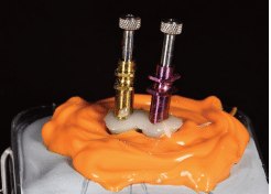
Once the material was dry, flowable resin-based composite was placed in the impression and cured. This step replicated the established tissue support (Figure 8). This was then removed and placed in the patient’s mouth for final impressions (Figure 9).
The porcelain veneers were seated with a flowable hybrid resin, and the three crowns were seated with a luting agent.
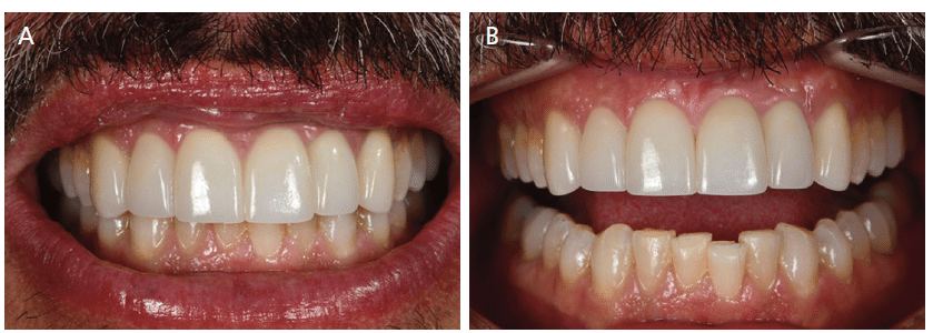
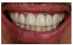
FINAL OUTCOME
Figures 10A and 10B show unretracted and retracted views of the finished case. Following almost eight months of restorative treatment, the patient was pleased with the results and feels he has the nicest smile he has ever had in his entire life.
American Academy of Cosmetic Dentistry
800-543-9220
From Decisions in Dentistry. March 2023;9(3)24-25.
