
Clinical Considerations for Delivery of Immediate Dentures
An exploration of the advantages of immediate dentures compared to conventional complete denture treatment.
PURCHASE COURSE
This course was published in the December 2019 issue and expires December 2022. The author has no commercial conflicts of interest to disclose. This 2 credit hour self-study activity is electronically mediated.
This course was published in the December 2019 issue and expires December 2022. The author has no commercial conflicts of interest to disclose. This 2 credit hour self-study activity is electronically mediated.
EDUCATIONAL OBJECTIVES
After reading this course, the participant should be able to:
- Discuss treatment planning and delivery of immediate dentures.
- List the advantages and disadvantages of immediate denture treatment.
- Explain clinical considerations pertaining to technique and patient follow-up in this approach to care.
Providing removable prosthodontic treatment is a key function in restorative dentistry. This facet of care will become increasingly common as the number of partially dentate and completely edentulous individuals rises due to increased life expectancy. Some of these patients will encounter tooth loss as a result of decay, trauma or periodontal disease, and dental clinicians should be able to meet these patients’ needs with quality removable prosthodontic treatment. One service that is commonly needed is an immediate denture if one or several teeth require removal. Though well documented in the literature,1–21 this procedure and the steps leading up to the extraction and prosthesis delivery appointment can be accomplished with minimal complications.
An immediate denture is a removable prosthesis that is placed at the time of extraction of a few or most of the dentition in one or both arches.22 First introduced and discussed by Jaffe23 and Hooper24 in the 1930s, the concept of immediate complete denture has undergone little significant change since then — that is, with the major exception of the advent of digital scanning and computer aided design/computer aided manufacturing (CAD/CAM) technologies.
There are a number of advantages7,25,26 to immediate dentures compared to conventional complete denture treatment. Once inserted, the denture base acts to control hemorrhage, maintain the position of the blood clot, and helps minimize swelling. It protects the surgical area (like a “plastic bandage”) from trauma or insult caused by opposing teeth, oral fluids and food debris. In addition, the transition from having some teeth to total edentulism is easier for the patient because facial tone is maintained, vertical dimension is not compromised, and speech patterns are minimally affected. Having a denture placed at the time of extraction has psychologically and financially benefitted those individuals who could ill afford to be out of the public eye for any prolonged time due to business or social demands. By having only anterior teeth remaining during denture construction, the clinician can utilize a record base and occlusion rim to better stabilize a face bow transfer, ensure a uniform occlusal plane, establish vertical dimension of occlusion, obtain and verify the centric relation record, and provide the patient with an esthetic try-in of the posterior teeth.
vertical dimension due to lack of posterior occlusion.
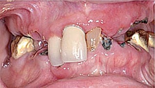
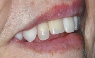
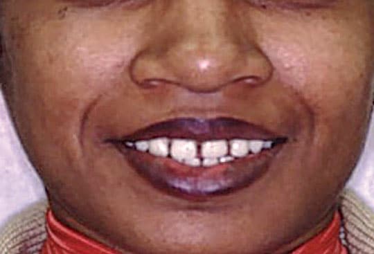
Disadvantages of this technique include prolonged treatment, increased expense due to additional procedures, impaired chewing efficiency from the lack of posterior teeth, and the potential alteration of speech and swallowing if there were alterations of tooth position. Additionally, the dentures cannot be fully assessed until insertion. Major adjustments and addition of reline material may be necessary at the delivery appointment. There may be circumstances in which not reproducing the original natural tooth position would provide more desirable esthetics. This scenario should be evaluated during the examination and discussed with the patient in regard to possible outcomes if artificial teeth are moved into a more favorable alignment — not only for esthetics, but also for function and speech (Figures 1A through 1C). Additionally, when planning for an immediate denture in the maxillary arch, the opposing arch must be evaluated to determine the need for occlusal plane correction or providing posterior occlusion if the patient is missing molars and/or premolars. Stability and retention of the maxillary denture is improved if opposing posterior dentition is present. Contraindications for this technique include patients who are medically compromised, psychologically unstable or who require daily nursing care.

After a thorough examination of the patient and discussion of the medical history, the dentist must ensure no treatment contraindications exist and the patient is medically stable to undergo multiple extractions with minimal risk. The patient’s expectations regarding immediate denture therapy should be discussed; sometimes, visual aids using models or pictures will help when explaining procedures. As part of case planning, patients should be made aware of the detailed aspects of immediate denture treatment. This is important because the process can be a traumatic introduction to removable prostheses if patients are not informed about the need for maintenance and the additional expense associated with treatment.
The importance of several months of continuing care for these patients must be explained, and the drawbacks of neglecting continuing care should be emphasized.12 Following insertion of an immediate denture, the patient may require a reline after a healing period of a few weeks. If significant changes to the ridge resulting from the delivery have caused instability of the prosthesis, the denture will require a soft reline. In light of this possibility, some authors advocate that immediate dentures should be considered as “treatment dentures” that should eventually be replaced with another set using more conventional prosthodontic principles. However, the added expense of a second set may be a deterrent to those on fixed incomes.27–29
SERIAL VERSUS COMPLETE ARCH EXTRACTIONS
There are valid reasons to consider planning a serial or two-phase extraction regimen. With serial extractions, the posterior teeth are removed and these sites are allowed to heal for six to eight weeks. This technique provides a more stable basal seat for the denture. It also can lessen the chance that a complete refabrication of the prosthesis will be necessary because the stability of the denture base is more predictable.
In cases involving complete extraction and simultaneous delivery, clinicians are challenged by the difficulty of obtaining an accurate initial impression due to the presence of teeth. This adds unpredictability regarding estimation of the final anatomy of the remaining ridge and associated soft tissues. Clinically, there is greater potential for post-insertion problems, such as adjustments to correct occlusal discrepancies and managing instability of the prosthesis. As such, ridge contours should be evaluated prior to surgery. For example, bony undercuts pose a problem with path of insertion and patient comfort if not addressed when anterior extractions are performed. Using a dental surveyor to study contours on diagnostic casts has been advised for different treatment procedures under specific circumstances associated with the depth and size of the undercut. As is the case for all procedures, detailed planning can lead to more predictable and successful outcomes.30
FINAL IMPRESSION TECHNIQUE
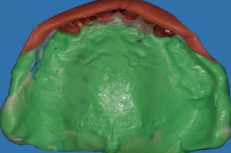
To adequately reproduce the anatomy, the final impression must produce a working cast with proper border extension and no distortion; it must also contain the maximum basal seat area. Methods to obtain such a working cast include using a stock or custom tray with irreversible hydrocolloid, vinyl polysiloxane, or some combination using split, custom impression trays or a hybrid technique.12,31,32 Regardless of the method utilized, the resulting impression must accurately reproduce the patient’s anatomy. Irreversible hydrocolloid in a stock tray that overlays a custom impression tray has been a standard procedure to achieve a detailed final impression for an immediate complete denture. Another method is to use a sectional impression technique in which the border molded custom impression tray captured most of the edentulous area distal to the natural teeth, followed by a putty matrix that captures the labial aspects of the remaining teeth, along with the labial vestibule.33,34 Since it is in two pieces, it can easily be removed without much discomfort for the patient if mobile teeth or severe tissue undercuts are present (Figures 2A through 2C). Once removed, the impression is reassembled and stabilized in order to make the final working cast from vacuum-mixed type III dental stone.
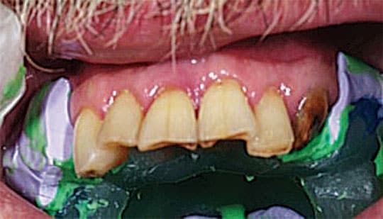
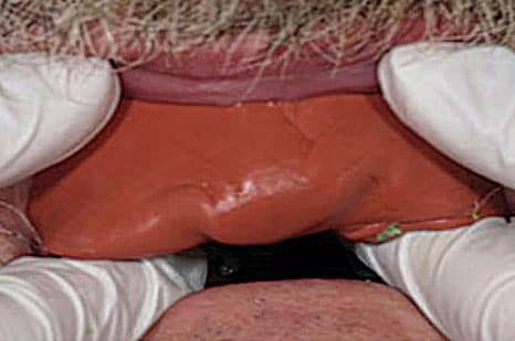
putty matrix and indexed to a custom
impression tray.
In order to determine the proper maxillomandibular relationship for the patient, having some natural teeth remaining can be helpful in establishing the vertical dimension of occlusion. Techniques commonly used in complete denture therapy will be useful when evaluating anterior tooth position to help determine proper lip support, phonetics and occlusal plane. Record bases and occlusion rims fabricated from the master cast aid in facebow transfer and making centric jaw relation records in order to transfer the relationship to the articulator for tooth set-up.
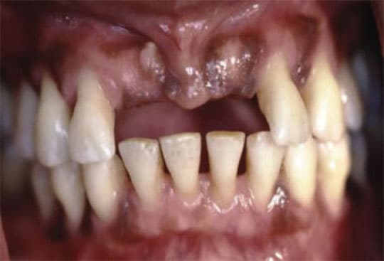
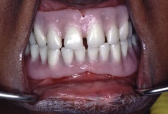
Discussions with the patient regarding anterior tooth arrangement are key to ensuring a desirable yet functional prosthesis (Figures 3A through 3C). Most of the time the teeth are reset in close proximity to the original relationship. However, as a result of advanced periodontal disease, some patients present with flared or extruded teeth that create an undesirable appearance. Previous photographs or casts will prove helpful in achieving a more attractive tooth position. It is not uncommon to invite the patient for an additional appointment before extractions to preview the final arrangement in order to gain approval before the denture is processed.
Modification of the cast with the removal of teeth should be done in a step-wise manner to aid in positioning of the denture teeth, as well as preserve and maintain ridge contour. Jerbi35 discusses a technique in which the labial ridge on the cast is divided into three equal zones, from the gingival margin to the vestibular roll. Once the clinical crown is removed from the cast, the labial portion of the stone cast is removed in a continual diminishing depth as it approaches the vestibule. Others suggest this method of cast modification is too aggressive and that minimal adjustment to the stone model will suffice.36,37
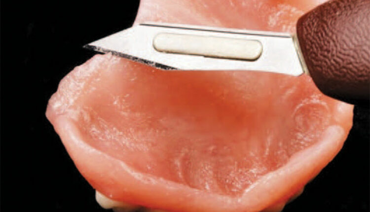
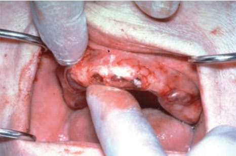
In order to achieve the desired shape of the alveolar ridge, a surgical template38–40 or matrix can be utilized at the time of surgery. This will help determine the need for bone recontouring associated with unwanted undercuts that would otherwise be problematic for the patient in terms of denture insertion and removal. The template can be fabricated from a duplicate of the working cast using clear thermoplastic material or autopolymerizing clear poly methyl methacrylate. At the time of extraction, the clear template is placed to check for tissue blanching. This allows the surgeon to detect areas that require recontouring and make adjustments to the alveolus before inserting the immediate prosthesis (Figure 4).
Inclusion of a labial flange to the maxillary immediate denture has been subject to debate. Some clinicians believe that if the anterior contours are severe, the flange is unnecessary or should be minimal since it could create too much distortion of the lip or irritation to the ridge. If needed, this structure can be added to the maxillary denture base using autopolymerizing denture resin as the patient progresses through normal healing.7,41
Following extractions, any needed alveoloplasty, and insertion, the patient should be directed to wear the prosthesis continually during the first 24 to 48 hours. The majority of swelling occurs during this time. Patients who receive immediate dentures may experience adverse physiologic and psychological effects, which may be caused by ill-fitting dentures that result from oral changes after insertion. To improve comfort and acceptance, written instructions should be given to each patient.42 The patient should be seen during the first 48 hours after the procedure to evaluate fit and address any pressure areas due to the denture base or flange extension. Minor occlusal adjustment may be necessary. Following the initial procedure, the patient should be checked again in approximately seven to 10 days. Because recontouring and healing tend to progress slowly and may stabilize as much as six to eight weeks later, the patient should be periodically evaluated to make any needed adjustments to enhance fit and ensure proper occlusion. Some type of intermediate reline material can be used to stabilize and aid retention (Figure 5).
As the technology associated with digital impressions43–45 and CAD/CAM prosthetics continues to improve, procedures such as immediate denture fabrication can become more streamlined and less invasive. Direct scanning of the entire arch can be achieved with great accuracy. The images captured using this modality, along with conventional photography, can help create a denture that is both esthetic and well fitting. Perhaps one day this will become the standard of care for removable prosthetics and replace the conventional therapy currently used.

CONCLUSION
Immediate denture treatment can be highly successful with proper planning, attention to detail, and good patient/provider communication. An explanation of the related procedures and how they are sequenced can be beneficial to patients as they transition to a state of edentulism. A stable foundation can be achieved with serial extractions and contouring the posterior ridges to support the denture base. Using modified custom impression trays, the final impression helps operators achieve the desired working cast.
Obtaining a patient-appropriate vertical dimension and centric jaw relation is no different than in a standard complete denture prosthodontic technique. As digital technology improves, treatment will become more streamlined, with less chairtime for both the clinician and patient. Gaining patient trust through communication during case planning and treatment will help minimize complications and support successful outcomes. Finally, the patient should be made aware that maintaining good oral hygiene and following the dentist’s recommendations will help minimize soft tissue irritation and ensure proper function and satisfaction.
REFERENCES
- Khan Z, Haeberle CB. One-appointment construction of an immediate transitional complete denture using visible light-cured resin. J Prosthet Dent. 1992;68:500–502.
- Haeberle CB, Khan Z. Construction of a custom-shaded interim denture using visible-light-cured resin. J Prosthodont. 1997;6:153–156.
- Woloch MM. Nontraumatic immediate complete denture placement: a clinical report. J Prosthet Dent. 1998;80:391–393.
- Harvey WL. A transitional prosthetic appliance. J Prosthet Dent. 1964;14:60–70.
- Johnson K. A clinical evaluation of upper immediate denture procedures. J Prosthet Dent. 1966;16:799–810.
- Rayson JH, Wesley RC. An intermediate denture technique. J Prosthet Dent.1970;23:456–463.
- LaVere AM, Krol AJ. Immediate denture service. J Prosthet Dent.1973;29:10–15.
- Swoope CC, Depew TE, Wisman LJ, Wands DH. Interim dentures. J Prosthet Dent.1974; 32:604–612.
- Herman GL. Esthetic and emotional factors in immediate denture construction. Compend.1989;10:486–488.
- Joffe EH. Simplified fabrication of the interim denture using a vacuum-forming machine: a clinical report. J Prosthet Dent.1992;67:747–748.
- Raczka TC, Esposito SJ. The “jiffy” denture: a simple solution to a sometimes difficult problem. Compend Contin Educ Dent.1995;16:914–18.
- Seals RR, Kuebker WA, Stewart KL. Immediate complete dentures. Dent Clin North Am. 1996;40:151–167.
- Duncan JD, Stephens AP, Caughman WF. Reproducing former esthetic qualities in new dentures. J Prosthet Dent.1988;60:515–517.
- Livaditis GJ. Indexing procedures for converting removable partial dentures after extractions while the patient retains the prosthesis. J Prosthet Dent.1999;81:485–491.
- Mou SH, Chai T. The pontic-splinted procedure for tooth and denture base additions in denture repair. J Prosthet Dent. 2001;85:126–128.
- Bouma LO, Mansueto MA, Koeppen RG. A nontraditional technique for obtaining optimal esthetics for an immediate denture: a clinical report. J Prosthodont. 2001;10:97–101.
- Sadowsky SJ, Gupta S, Gonzales E. A technique to correct incisal plane error in maxillary immediate denture therapy. J Prosthet Dent. 2013;110:141–143.
- Lee J. Fabricating an immediate denture for a medically compromised elderly patient. J Prosthet Dent. 2015;113:277–281.
- Gotlieb AS, Askinas SW. An atypical chairside immediate denture: a clinical report. J Prosthet Dent. 2001;86:241–243.
- Gooya A, Ejlali M, Adli AR. Fabricating an interim immediate partial denture in one appointment (modified jiffy denture). A clinical report. J Prosthodont. 2013;22:330–333.
- Michalakis KX, Touloumi F, Calvani L, Bedi A, Hirayama H. Simplifying prosthetic procedures while converting an interim maxillary removable complete denture to an interim implant-supported fixed complete denture. J Prosthodont. 2011;20:408–413.
- Glossary of Prosthodontic Terms, 9th ed. Available at: https://www.academyofprosthodontics.org/_Library/ap_articles_download/GPT9.pdf. Accessed September 26, 2019.
- Jaffe SS. Immediate full restorations. J Am Dent Assoc. 1932;19:396.
- Hooper JL. Immediate denture technique insuring preservation of facial dimension. Dent Dig. 1937;43:475.
- Bruce RW. Immediate denture service designed to preserve oral structures. J Prosthet Dent. 1966;16:811–821.
- Rahn AO, Ivanhoe JR, Plummer KD. Textbook of Complete Dentures. 6th ed. Shelton,CT: People’s Medical Publishing House;2009:272.
- Pound E. Controlled immediate dentures. J South Calif Dent Assoc. 1970;38:810–817.
- Payne S. A transitional denture. J Prosthet Dent. 1964;14:221–230.
- Clark RK, Radford DR. Immediate replacement complete dentures: pitfalls of ignoring traditional teaching and established practice. Eur J Prosthodont Restor Dent. 2011;19:131–134.
- Demer W. Minimizing problems in placement of immediate dentures. J Prosthet Dent.1972;27:275–284.
- Beumont AJ. Sectional impression for maxillary Class I removable partial dentures and maxillary immediate dentures. J Prosthet Dent.1983;49:438–441.
- Lee H, Park C. A method to make a preliminary impression of mobile teeth. J Prosthet Dent. 2009;102:52–53.
- Gardner LK, Parr GR, Rahn AO. Modification of immediate denture sectional impression technique using vinyl polysiloxane. J Prosthet Dent. 1990;64:182–184.
- Cagna DR, Massad JJ. Vinyl polysiloxane impression material in removable prosthodontics. Part 2: immediate denture and reline impressions. Dent Clin North Am. 2007;28:519–526.
- Jerbi FC. Trimming the cast in the construction of immediate dentures. J Prosthet Dent. 1966;16:1047–1053.
- Standard SG. Preparation of casts for immediate dentures. J Prosthet Dent. 1958;8:26–30.
- Phoenix RD, Fleigel JD. Cast modification for immediate complete dentures: traditional and contemporary considerations with an introduction of spatial modeling. J Prosthet Dent. 2008;100:399–405.
- Bissasu M. A simple procedure for minimizing adjustment of immediate complete denture: a clinical report. J Prosthet Dent. 2004;92:125-127.
- Gilboa I, Cardash HS. An alternative approach to the immediate overdenture. J Prosthodont. 2009;18:71–75.
- Farmer JB. Surgical template fabrication for immediate dentures. J Prosthet Dent.1983;49:579–580.
- Pound E. Achieving patient acceptance to immediate denture service. J Am Dent Assoc. 1963;66:795–802.
- Holt RA Jr. Instructions for patients who receive immediate dentures. J Am Dent Assoc. 1986;112:645–646.
- Versteeg, G. Immediate dentures based on digital impressions and digital workflow. Available at: https://www.aegisdentalnetwork.com/special-issues/2019/08/immediate-dentures-based-on-digital-impressions-and-digital-workflow. Accessed September 26, 2019.
- Neumeier TT, Neumeier H. Digital immediate dentures treatment: a clinical report of two patients. J Prosthet Dent. 2016;116:314–319.
- Goodacre CJ, Garbacea A, Naylor WP, Daher T, Marchack CB, Lowry J. CAD/CAM fabricated complete dentures: concepts and clinical methods of obtaining required morphological data. J Prosthet Dent. 2012;107:34–46.
From Decisions in Dentistry. November/December 2019;5(10):36,39—41.

