

The Small Canal Behind Big Endodontic Failures
Mastering the diagnosis and management of hidden anatomy requires the right combination of three-dimensional imaging, enhanced visualization, and minimally invasive techniques.
One of the most common reasons for endodontic retreatment is the discovery of a previously missed MB2 canal. Although clinicians have more diagnostic and treatment technologies than ever before, untreated canals continue to significantly contribute to endodontic failure in maxillary molars.
Research consistently demonstrates a strong relationship between missed canals and unsuccessful root canal therapy.1 The objective of both primary treatment and retreatment is complete elimination of infection within the root canal system while preserving the natural tooth. Failure to identify all canals, whether due to anatomical complexity, limited visibility, inadequate knowledge of internal anatomy, or underutilization of available technology, can greatly reduce the long-term prognosis.2
Endodontic retreatment is often performed to manage failed primary root canal therapy, but its success rate of 75% to 85% falls well below the 94% success rate reported for primary root canal treatment (RCT).3 Incomplete RCT due to the lack of canal localization and therapy, difficulty in internal anatomy management, bacterial leakage in the marginal gap during the final restorations, delayed restoration, and new decay on the tooth structure are some of the main reasons for treatment failure.4
Research shows the overall prevalence of missed canals among endodontically treated teeth is approximately 13% to 23.4%.5-7 The prevalence of missed canals is higher in maxillary first molars (40.6%). The overall prevalence of apical periodontitis among teeth with missed canals is approximately 90%. The second mesiobuccal canal in the maxillary first molars and the mesiobuccal and distobuccal canals in mandibular teeth are the most commonly missed.2 Failure to locate and treat all root canals results in inadequate mechanical debridement and persistent bacterial infection, increasing the risk of apical periodontitis and its associated clinical signs, including inflammation, pain, sinus tract formation, and periapical radiolucency.4
Adequate microbiology and internal anatomy control are key factors in RCT. Technologies, such as cone-beam computed tomography (CBCT), dental microscope, and ultrasonic scaler, are fundamental for successful RCT and retreatment. The three-dimensional images provided by CBCT offer multiple views to detect the rarefaction’s size, position, and extension. They also help detect failures, such as incomplete treatment, internal or external resorptions, and missed canals.8,9
Importance of Visualization
High-quality endodontic care depends on enhanced visualization of the complex root canal system. The use of the dental microscope is the standard of care in multidisciplinary dentistry. In endodontics, it is absolutely necessary to perform precision tasks in deep, narrow, and dark spaces such as the pulp chamber and root canals.10
A clinician’s hands become more skilled as visual capacity increases. Compared with unaided vision or low-power (2×) loupes, an operating microscope provides substantially greater magnification and dramatically improved resolution, allowing clinicians to distinguish fine anatomical details with far greater accuracy.11 Research shows the importance of magnification in the management of canals’ internal anatomy, especially when locating the MB2 canal of the first and second molars, which is one of the most challenging procedures in complex endodontics.12
One of the greatest advantages of the dental microscope in endodontics is its ability to improve the detection of complex root canal anatomy, including the MB2 canal. Compared with unaided vision, the combination of variable magnification and coaxial LED illumination provides exceptional visualization of the operating field, enabling clinicians to identify anatomical details that might otherwise be missed. Enhanced visualization not only improves treatment precision but also promotes better ergonomics by allowing clinicians to maintain a more upright, neutral working posture.13 The dental microscope and ultrasonic scaler are critical parts of the endodontist’s armamentarium. The combination of enhanced magnification and coaxial illumination provided by the dental microscope, together with the slim diameter, angulation, and extended working length of ultrasonic tips, allows clinicians to conservatively remove dentin and canal obstructions while maintaining clear visualization of the deepest portions of the root canal system.14
Locating the MB2 canal in maxillary molars begins with careful visual examination of the pulp chamber floor under the dental microscope. Developmental map lines, combined with tactile exploration using an endodontic explorer between the mesiobuccal and palatal canal orifices, often guide the clinician to the MB2 canal, which is typically located 2 to 3 mm palatal and slightly mesial to the MB1 canal.15 Additional visual clues, including the white line test, red line test, presence of moisture at the canal orifice, and the bubble test, can further aid in identifying this elusive canal.16
Selective RCT is an option in endodontic retreatment cases when periapical pathosis is present in a specific root canal in a multirooted tooth while the other periapical tissue remains healthy.17 A valuable resource, selective RCT needs to be explained to the patient to ensure full understanding and informed acceptance. The recommendation should be based on key diagnostic and prognostic factors, including the extent of any required crown or restoration adjustments, the quality and integrity of the root canal filling following RCT, and the presence or absence of periapical pathology identified on radiographic examination. The final decision should be made after the dentist has accessed the tooth and analyzed internal factors in the pulp chamber, such as the degree of contamination or filling quality in the other root canals. A selective RCT may reduce time and costs for the patient and should be considered in appropriate cases.
Case Report
A 50-year-old woman was referred to the office for a consultation. The patient manifested mild spontaneous pain and sensitivity when biting and touching tooth number three, with a history of a root canal treatment done 1 year ago. Clinically, the tooth presented a well-adjusted ceramic crown without mobility and inflammation in the soft tissues around the mesial root.
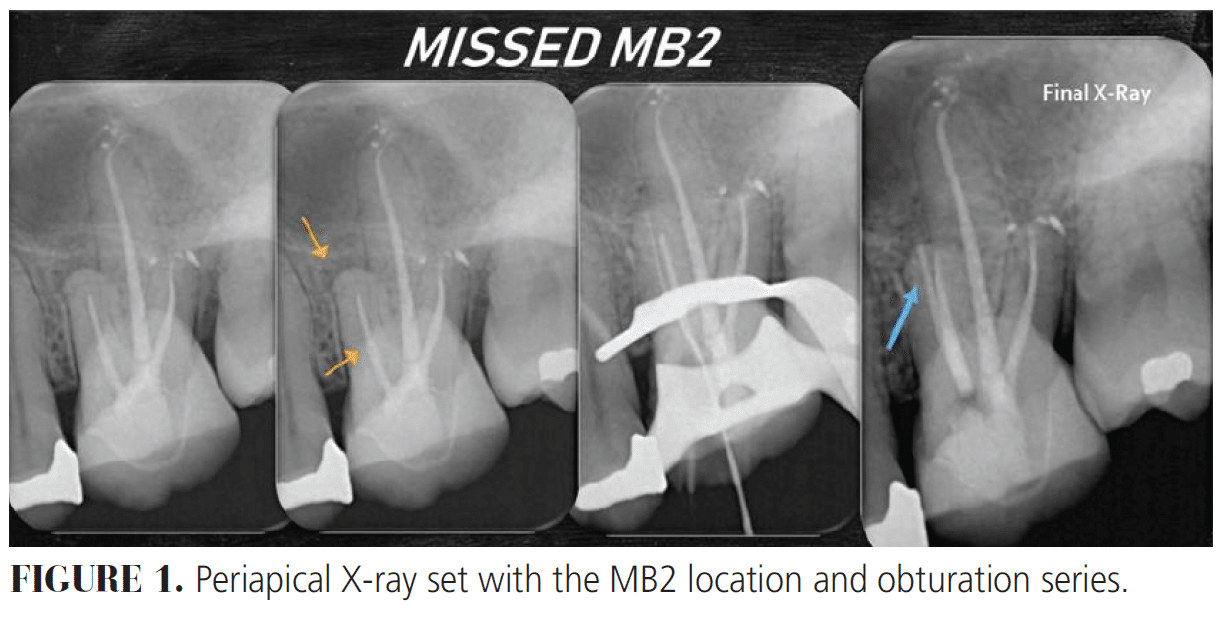
We took three different angulation X-rays. The mesial root was the only one with a periapical rarefaction; the two other root canals presented normal radiographical aspects in the periapical area. By analyzing the periapical X-ray, we considered that the mesial root canal filling was poor, manifesting a possible missed MB2 (Figure 1).
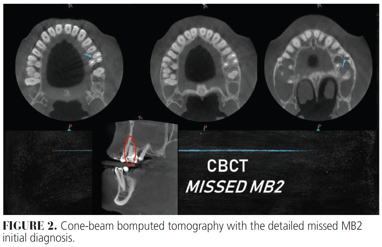
We suggested CBCT to gather all possible information to complete the diagnosis. By analyzing the CBCT scan in the axial plane, the missed MB2 was detected and the palatal and distal roots were shown to be healthy (Figure 2).
A selective mesial RCT was recommended due to the well-adjusted crown, healthy periapical tissues, and correct root canal filling on the palatal and distal roots. The endodontic access was performed under a dental microscope using low-to-medium magnification levels (4×, 6×, and 10×). A number two carbide bur was utilized to create the access, allowing for conservative removal of tooth structure with minimal alteration or wear of the existing crown and restoration (Figure 3A). When getting deeper into the pulp chamber floor, we switched to an ultrasonic scaler diamond bur tip to enable clear, deep vision into the internal area of the pulp chamber. The guttapercha was discovered when removing all the composite.
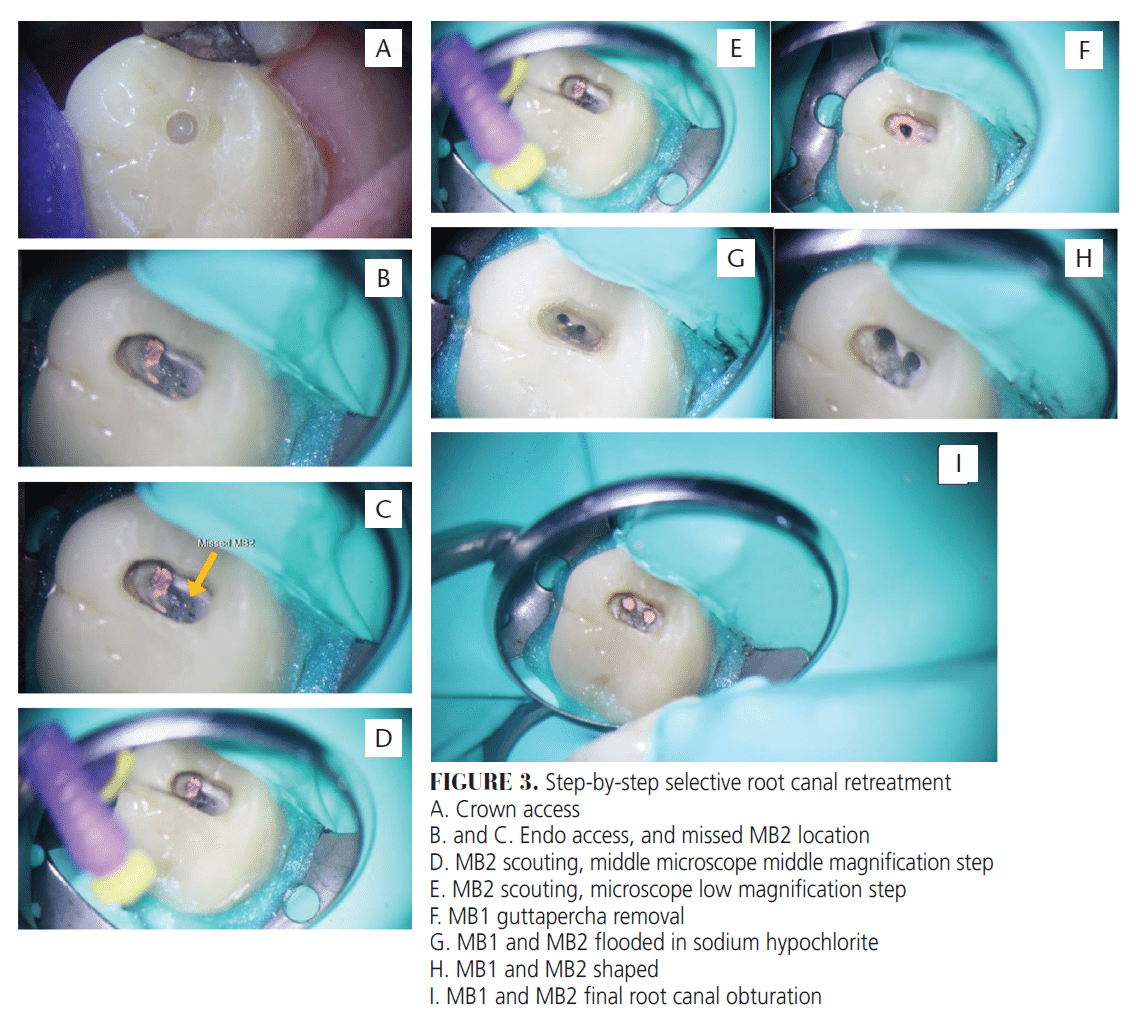
An E5 ultrasonic tip was used to conservatively trough dentin distally and palatally from the MB1 canal while adhering to the pulpal floor map and white line guidelines for MB2 canal identification. Ethylenediaminetetraacetic acid (EDTA) was periodically applied to maintain a hydrated pulpal floor, enhancing the visualization of anatomical color variations, texture changes, and developmental grooves. Under medium and high magnification with the dental microscope, progressive refinement of the access was performed until the MB2 canal orifice was identified by the characteristic humid bubbling pattern (Figure 3B and C).
Once the MB2 canal was located, we gauged and scouted the canal with #8, #10, and #15 K files on the cervical root third, recapitulating and constantly flooding the pulp chamber with sodium hypoclorite 5.25% between the K files. Then, we used an orifice shaper instrument to smooth the cervical dentin triangle for better access into the apical third of the root canal and a #10 K file in oscillatory motion with an oscillatory contra angle in 60° motion activated with the low handpiece (Figure 3D and E).
Once reaching the apical third, an apex locator with a teflon #15 file and periapical X-ray were used to confirm the working length. The step-back shaping technique was implemented. First, K file instrumentation and recapitulation were used to maintain the glide path with a #10 file until reaching a #35 K file to working length. Rotary files were used to shape the canal until reaching file 25.06, always using EDTA with every instrument from the sequence.
When recapitulating, passive ultrasonic irrigation was used by activating a K file #10 to the working length with the E5 ultrasonic tip for 10 seconds. Once reaching the apical third canal shape, we removed the MB1 guttapercha, using first an E5 ultrasonic tip to heat the guttapercha, followed by a endodontic rotary orifice opener; Hedstrom file #30, #35; and rotary files until reaching the approximate working length (WL). A #15 K file was then used to confirm the WL with an apex locator and periapical X-ray (Figure 3F).
The working length was confirmed, and the canal was shaped with rotary files, irrigated with sodium hydroxide and EDTA 17%, and finalized with a #35 K file. The sodium hypochlorite irrigation was activated with a #10 file by ultrasound, the final rinse was done with isopropyl alcohol, and paper points were used to dry (Figure 3G). After finishing the MB1 canal retreatment, the Tagger hybrid obturation technique was performed using the McSpadden Guttacondensor #35 in each canal, and then a composite filling was placed on top (Figure 3H and I).
The patient was scheduled for radiographic follow-up at 3 months to evaluate the healing response, monitor the repair process, and assess clinical progression.
Conclusion
Treating a first or second upper molar brings anatomical management challenges. Finding the MB2 canal is an absolute must. CBCT provides the clinician with a comprehensive understanding of the tooth’s internal anatomy, allowing evaluation of previous treatments, identification of potential causes of RCT failure, and development of a more predictable treatment strategy. This 3D assessment enhances diagnostic accuracy and provides greater confidence when approaching complex endodontic procedures.
Successful management of the MB2 canal requires a thorough understanding of root canal anatomy, including its typical location, anatomical variations, and clinical guidelines for identification, negotiation, cleaning, and shaping. The clinician must recognize the anatomical landscape and apply contemporary techniques to improve the probability of successful outcomes.
The identification and management of the MB2 canal is a highly demanding procedure that requires exceptional visual control and precision. Accurate dentin removal, avoidance of perforation risks, and recognition of subtle differences in color and texture require the use of multiple magnification levels under a dental microscope. Coaxial illumination provided by the microscope enhances visualization of anatomical details, allowing the clinician to distinguish critical structural variations and perform conservative dentin removal with improved control. Ultrasonic tips further contribute to the precision and safety of this delicate step.
A well-established technique for MB2 canal scouting and shaping is essential to achieve proper glide path development. The presence of a pronounced cervical dentin triangle may obstruct access to the remaining canal anatomy and compromise cleaning and shaping procedures. Careful removal of restrictive dentin, combined with the controlled use of rotary and/or reciprocating instruments and advanced irrigation protocols, facilitates safe and effective MB2 canal management.
This case report demonstrates the decision-making process involved in determining whether all root canals should be addressed during endodontic retreatment. It also highlights how technological advancements, appropriate diagnostic assessment, and a simplified, evidence-based retreatment protocol can help overcome the technical challenges associated with previously treated root canal systems.
References
- Gulabivala K, Ng YL. Factors that affect the outcomes of root canal treatment and retreatment — a reframing of the principles. Int Endod J. 2023;56(Suppl 2):82-115.
- Mashyakhy M, Hadi FA, Alhazmi HA, et al. Prevalence of missed canals and their association with apical periodontitis in posterior endodontically treated teeth: a CBCT study. Int J Dent. 2021;2021:9962429.
- Sabeti M, Chung YJ, Aghamohammadi N, Khansari A, Pakzad R, Azarpazhooh A. Outcome of contemporary nonsurgical endodontic retreatment: a systematic review of randomized controlled trials and cohort studies. J Endod. 2024;50:414-433.
- Tabassum S, Khan FR. Failure of endodontic treatment: the usual suspects. Eur J Dent. 2016;10:144–147.
- Rouhani A, Aboutorabzadeh SM, Reyhani M, Kheirabadi N, Mortazavi S, Navabi S. Prevalence of missed canals in endodontically treated teeth: A cone-beam computed tomography study. J Clin Exp Dent. 2023;15:e605–e611.
- Costa F, Pacheco-Yanes J, Siqueira Jr J, Oliveira A, Gazzaneo I, Amorim C. Association between missed canals and apical periodontitis. J Endod. 2019;52:400-406.
- Karabucak B, Bunes A, Chehoud C, Kohli MR, Setzer F. Prevalence of apical periodontitis in endodontically treated premolars and molars with untreated canal: a cone-beam computed tomography study. J Endod. 2016;42:538-541.
- Venkatesh E, Elluru SV. Cone beam computed tomography: basics and applications in dentistry. J Istanb Univ Fac Dent. 2017;51(3 Suppl 1):S102–S121.
- Scarfe WC, Levin MD, Gane D, Farman AG.Use of cone beam computed tomography in endodontics. Int J Dent. 2010;2009:634567.
- Carr GB, Murgel CAF. The use of the operating microscope in endodontics. Dent Clin North Am. 2010;54:191-214.
- van As GA. Magnification alternatives: seeing is believing, Part I. Dentistry Today. 2013;32(6):82-87.
- Alaçam T, Tinaz AC, Genç O, Kayaoglu G. Second mesiobuccal canal detection in maxillary first molars using microscopy and ultrasonics. Aust Endod J. 2008;34:106-109.
- Buhrley LJ, Barrows MJ, BeGole EA, Wenckusl CS. Effect of magnification on locating the MB2 canal in maxillary molars. J Endod. 2002;28:324-327.
- Sujith R, Dhananjaya K, Chaurasia VR, Kasigari D, Veerabhadrappa AC, Naik S. Microscope magnification and ultrasonic precision guidance for location and negotiation of second mesiobuccal canal: An in vivo study. J Int Soc Prev Community Dent. 2014;4(Suppl 3):S209–S212.
- Coelho MS, Lacerda MFLS, Silva MHC, Rios MA. Locating the second mesiobuccal canal in maxillary molars: challenges and solutions. Clin Cosmet Investig Dent. 2018;10:195–202.
- Mulay S, Kadam G, Jain H. Accuracy of various diagnostic aids in detection of mb2 canal in maxillary first molar: in vivo study. World Journal of Dentistry. 2016;7(2):78-82.
- Nudera WJ. Selective root retreatment: a novel approach. J Endod. 2015;41:1382-1388.
From Decisions in Dentistry. September/October 2026;12(2):10-14.