
 ALEX-MIT/ISTOCK/GETTY IMAGES PLUS
Modern Applications for The Cantilever Fixed Partial Denture
As this case report demonstrates, certain clinical indications suggest the use of a cantilever fixed partial denture.
A cantilever fixed partial denture is defined as one that has an abutment or abutments at one end only, with the other end of the pontic remaining unattached.1 With the advent of single-tooth implants, use of the cantilever fixed partial denture has decreased over the years. When cantilevered partials are utilized, they most frequently replace a single tooth, such as a lateral incisor. Due to the added stress on the pontic and single abutment, careful attention must be paid to the occlusion, periodontal support, abutment crown length, and the crown-to-root ratio.2
A systematic review of cantilever fixed partial dentures reported an 18.2 % failure rate at 10 years, as compared to 10.9% with use of a conventional fixed partial denture.3 The most frequent complication was loss of retention, with the second most common failure being fracture of the restorative material.3 There are occasions, however, when a complicated medical history or financial constraints make a cantilever fixed partial denture a reasonable option, as opposed to an implant or conventional fixed partial denture. This article presents a case report where a cantilever fixed partial denture was utilized in lieu of either the implant or conventional fixed partial denture option. This case specifically involved replacing the maxillary canines bilaterally — both using a single premolar abutment.
Replacing a maxillary canine with a fixed partial denture is an especially challenging situation. The challenge lies in replacing one of the strongest teeth in the arch — and one typically significantly involved in occlusion — using the weakest teeth in the arch, the maxillary lateral incisor and maxillary first premolar, as abutments. The significant role canines play in occlusion poses a considerable challenge when considering restorative options. When considering a cantilevered restoration, using double abutments to address issues of retention, resistance and occlusion has been recommended.2,4 In the case presented here, after considering the patient’s medical history, status of abutment teeth, occlusion, financial situation and desires, the decision was made to use bilateral cantilever fixed partial dentures to replace maxillary canines utilizing a single premolar abutment.
CASE PRESENTATION
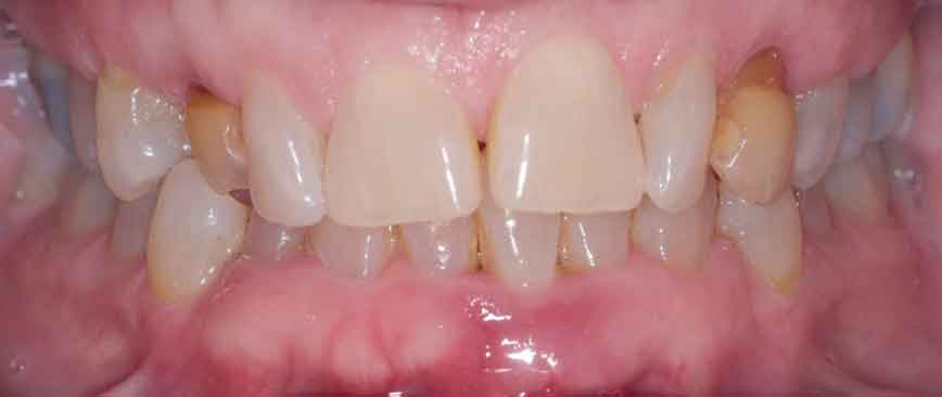
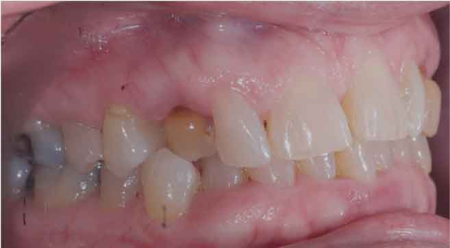
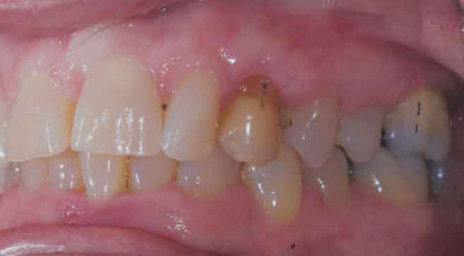
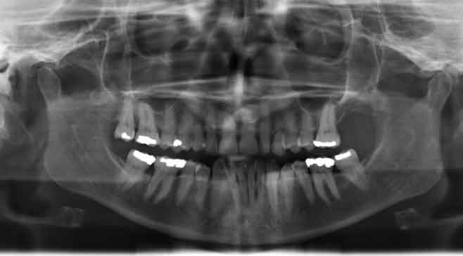
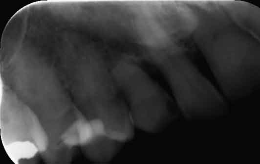
A 68-year-old female presented to the student clinic at the Dental College of Georgia at Augusta University with a chief complaint of wanting to replace two retained primary canines, teeth C and H. Both permanent maxillary canines (teeth #6 and 11) were horizontally impacted (Figure 1 through Figure 6B). Her medical history was positive for hypertension, hyperlipidemia, anxiety and osteopenia. The osteopenia was being treated with an oral bisphosphonate on a weekly basis. The patient had been on the medication for more than two years and was classified as ASA 2a (mild controlled disease) under the American Society of Anesthesiologists physical status classification system.

The patient started treatment in the student clinic in 2017 after complaining that her retained primary canines were “loose and discolored.” Her initial treatment consisted of periodontal therapy, along with consultation regarding the primary canines. She presented with mild to moderate periodontitis, with multiple sites having probing depths of 5 to 7 mm in the posterior quadrants. She also had localized severe periodontitis involving teeth #24 and 25, and around the retained primary canine H. Treatment consisted of scaling, root planing and oral hygiene reinforcement. An orthodontic consultation was also completed, but, due to the severity of the impactions and the patient’s age, that option was eliminated.
During a consultation in January 2018, the possibility of a fixed replacement option was initially discussed. At the periodontal reevaluation in April 2018, 5 mm probing depths were noted around the primary canines. Treatment options were again discussed, and the patient expressed concern that the “baby teeth looked dark and aged.” The patient continued with periodontal therapy and was assigned to the eventual student provider in June 2019.
The oral examination revealed primary caries, Class 3 mobility, and discoloration of teeth C and H. Secondary caries was noted on the distal of tooth #5, with primary caries on the mesial of #4. The patient presented with Class 1 occlusion bilaterally, with crossbite on the right side involving tooth C. She also had anterior guidance on her right excursion, and group function on left excursive movement. A treatment plan was developed which included cone beam computed tomography, consultations with periodontists and prosthodontists, diagnostic casts, and diagnostic waxing.
The consultations resulted in several possible treatment approaches. An implant supported crown would be the restoration of choice in many cases; however, implants were not recommended due to ankylosis of teeth #6 and 11, and long-term bisphosphonate use by the patient. A conventional fixed partial denture using the central incisors, lateral incisors and first premolars as abutments had also been considered, but the patient did not want to involve her maxillary incisors with a conventional fixed partial denture if possible. A three-unit cantilever fixed partial denture extending from the maxillary second premolar was another treatment option presented, along with the option of a fixed partial denture using the first premolars as single abutment retainers.
Preliminary treatment included continuation of the periodontal therapy initiated previously, as well as treatment for the caries on teeth #4 and 5. Probing depths were now noted to be 5 to 8 mm between teeth H and #12, and 4 to 5 mm around #5 and C. Scaling and root planing was completed where indicated, and a final restorative treatment plan was developed.

In this case report, a two-unit cantilever fixed partial denture utilizing the maxillary first premolars as abutments with cantilever pontics was chosen to replace the missing canines. The option of using the second premolar as a double abutment was also considered, as it would add to the support, resistance, and retention of the fixed partial denture. However, it comes at additional cost to the patient, as well as an increased challenge to maintain oral hygiene around the splinted abutments. It was felt that the plan to carefully manage the occlusal scheme would allow the use of the single abutment.
Full veneer retainers would be fabricated on the first premolars. Both teeth #5 and 12 exhibited desirable characteristics for an abutment, including adequate root length, favorable crown-to-root ratios (between 1:1 to 2:3), and a healthy periodontium. The occlusal scheme would be designed to maximize the maximum intercuspation contact on the abutments, with light contact on the pontic. The plan called for maintaining excursive contacts only on the abutments, with no contact planned for the pontics.
The pontics would be designed as a modified ridge lap, with maximum occlusogingival height to ensure a rigid prosthesis. Essentially, the plan was to slightly modify the patient’s existing occlusion to group function bilaterally, minimizing stress on the pontics and, consequently, on the abutments.
SEQUENCE OF TREATMENT
The primary canines were extracted in September 2019, but immediate provisionals were not utilized. Healing proceeded as expected, with no complications. The soft tissue exhibited normal tissue contours at a two-week follow-up. The patient’s periodontal status had improved considerably, with most posterior probing depths measuring 2 to 3 mm, with only a few measuring 4 mm. Probing depths on both premolar abutment teeth also improved, with no depths greater than 3 mm recorded. Tooth #12 was prepared to serve as a porcelain-fused-to-metal (PFM) abutment six weeks from the extraction date, utilizing a standard PFM preparation of at least 1.5 mm occlusal reduction, 1 mm axial reduction, and a 0.5 mm chamfer, except for the facial margin, which was prepared as a rounded shoulder approaching 1 mm.1 Margins were to be at, or slightly below, the free gingival margin. The facial margins were designed for maximum esthetics using a porcelain shoulder.
A provisional fixed partial denture was fabricated using bisacryl from the diagnostic waxing made earlier. The patient returned five days later to have the margins refined and make the final impression using conventional vinyl polysiloxane using a two-cord technique for margin isolation. The working cast was cross-mounted to an opposing cast that had been previously mounted using a facebow for final fabrication on an articulator. Tooth #5 was prepared three days later to serve as a PFM cantilever abutment as well. A provisional fixed partial denture was again fabricated using bisacryl from a diagnostic waxing made earlier. The preparation on tooth #5 was completed in November 2019.
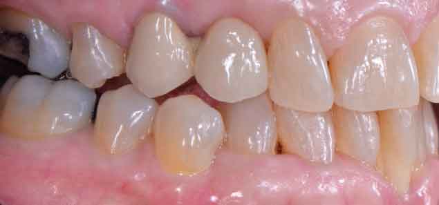
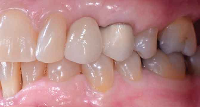
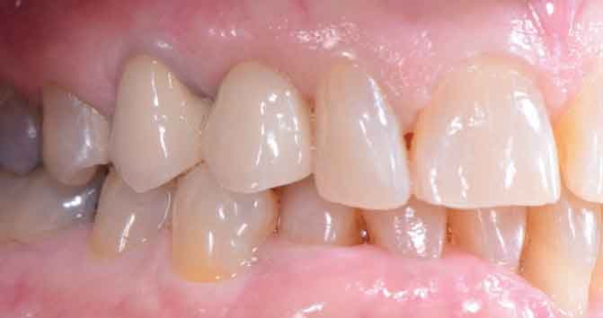
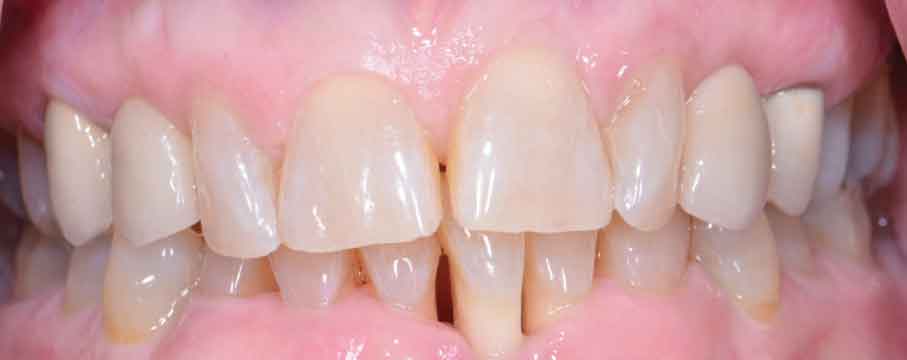
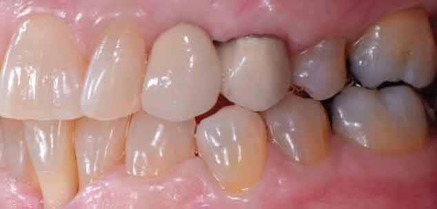
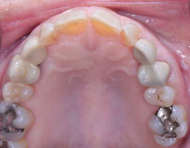
The fixed partial denture replacing tooth #11 was cemented two months later in January 2020 using a resin-modified glass ionomer luting cement. The preparation on tooth #5 was refined seven days after that, after which a final impression was made, and casts were again hand articulated and cross-mounted on an articulator for fabrication. The fixed partial denture replacing tooth #6 was cemented with a resin-modified glass ionomer luting cement in March 2020 (Figure 7 through Figure 9).
Cementation of the last fixed partial denture occurred on the day before the clinic closed due to the COVID-19 pandemic. The patient returned six months later in September 2020 to continue periodontal treatment. She then returned in December 2020 for periodontal maintenance and to reevaluate hygiene, occlusion, and overall health of the fixed partial denture.
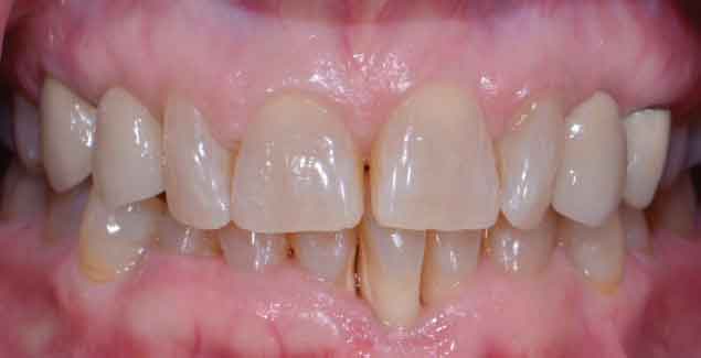
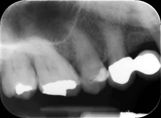
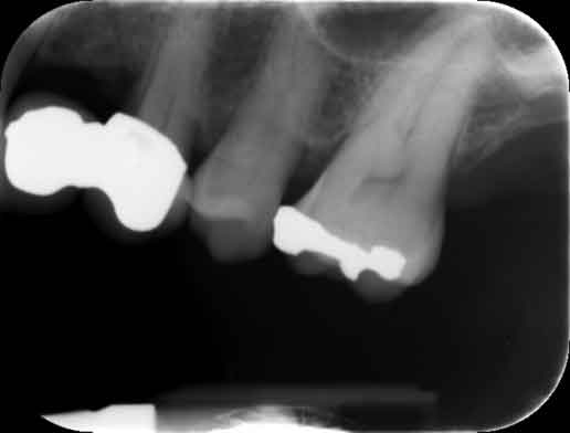
Treatment time from extractions to cementation was six months total. A final follow-up was completed in February 2021. This allowed a 13-month follow-up on the fixed partial denture replacing tooth #11, and 11 months for the fixed partial denture replacing #6 (Figure 10 through Figure 13). Figure 10 and Figure 11 both show early lateral excursive movements. Figures 14A and 14B and Figures 15A and 15B are post-cementation radiographs.
APPROACH TO TREATMENT
Managing the patient’s occlusion was paramount to a successful outcome in this case. The patient was missing both maxillary canines, teeth that are generally key components when developing an occlusal scheme — especially one involving anterior guidance. The plan to replace these key components with cantilever restorations required careful attention to the occlusal scheme to ensure a successful outcome for the single cantilevered abutments. The patient presented with group function on the right side, with heavy contact on the lateral incisors and light contact on the premolar. She also presented with group function on the left side. A limited occlusal adjustment was performed at the time of tooth preparation to lighten any excessively heavy excursive contacts, provide even contacts in maximum intercuspation, and minimize the impact of the crossbite on the right side by recontouring tooth #28. The planned occlusal scheme would place maximum intercuspation contacts on the abutments, with lateral excursions guided by either the abutments, lateral incisors, or both — essentially duplicating the patient’s existing occlusion. ‘
OUTCOME
The patient was extremely pleased with the esthetic results. She has maintained excellent hygiene around the fixed partial denture. Probing depths were noted to be less than 3 mm around the abutments, except for the mesial of tooth #12, which measured 4 mm. This was the area that initially presented with the greatest defect around the retained primary canine. Mobility on tooth #5 was noted to be within normal limits, while mobility on #12 appeared to be a 1. Evaluation of the occlusion in lateral excursions showed no guidance off the tooth #6 pontic, while the left lateral disclusion was shared by #10, as well as #11. The patient will be followed again in the student clinic and the plan will be to further minimize the forces on tooth #11 in lateral disclusion.
DISCUSSION
This case represents a departure from conventional guidance regarding the use of cantilever fixed partial dentures, which suggests using two abutments,1,2 particularly when replacing a maxillary canine. When faced with replacing a maxillary canine, the first option for many providers would be to utilize a single-tooth implant. The second option would likely be a conventional fixed partial denture utilizing the adjacent abutment teeth, possibly using double abutments. Due to the patient’s medical history, personal preferences, and existing occlusion, a third option was used for this patient — that of a cantilever PFM fixed partial denture utilizing the maxillary first premolars as the lone abutments. The fixed partial dentures were fabricated using standard preparations, as described earlier. Conventional noble alloys and feldspathic porcelain were also used. These materials provided the strength, rigidity and esthetics required in this case.

Long-term success in this case, as in all cases, will depend on the patient’s level of hygiene and maintenance of periodontal health.5 Regular maintenance visits will focus on these two factors, as well as the occlusal stability of the restorations and abutments. The patient had no history of bruxism, so an occlusal guard was not planned. The immediate esthetic results were excellent, and periodontal health, occlusion and mobility were healthy at the 12-month follow-up. Himmel et al’s4 review discusses 11 factors that lead to an improved prognosis when considering use of a cantilever fixed partial denture. Other than use of a double abutment, this case report demonstrates a successful outcome, at least in the short term, by successfully addressing the other criteria noted.

CONCLUSION
Effective treatment planning is crucial to successful outcomes. It is important to keep all options open when planning treatment, even some that are rarely utilized. In this case, the patient’s medical history, occlusion, oral hygiene, caries activity, personal preferences, and finances were all factors that played a significant role in the selection of the final treatment plan.
The authors have no commercial conflicts of interest to disclose.
KEY TAKEAWAYS
- A cantilever fixed partial denture is defined as one that has an abutment or abutments at one end only, with the other end of the pontic remaining unattached.1
- When cantilevered dental partials are utilized, they most frequently replace a single tooth, such as a lateral incisor.
- Optional restorative choices in these cases might include a single-tooth implant or conventional fixed partial denture.
- Due to the added stress on the pontic and single abutment, careful attention must be paid to the occlusion, periodontal support, abutment crown length, and the crown-to-root ratio.2
- This case report represents a departure from conventional guidance regarding the use of cantilever fixed partial dentures, which suggests using two abutments,1,2 particularly when replacing a maxillary canine.
- That noted, the patient’s circumstances dictated the approach outlined in this paper; namely, utilizing the maxillary first premolars as the lone abutments.
REFERENCES
- Shillingburg HT, Sather DA, Wilson Jr EL, et al. Fundamentals of Fixed Prosthodontics. 4th ed. Hanover Park, Ill: Quintessence Books; 2012:95.
- Wright WE. Success with the cantilever fixed partial denture. J Prosthet Dent. 1986;55:537–539.
- Pjetursson BE, Tan K, Lang NP, Brägger U, Egger M, Zwahlen M. A systematic review of the survival and complication rates of fixed partial dentures (FPDs) after an observation period of at least 5 years. IV. Cantilever or extension FPDs. Clin Oral Implants Res. 2005;15:667–676.
- Himmel R, Pilo R, Assif D, Aviv I. The cantilever fixed partial denture — a literature review. J Prosthet Dent. 1992;67:484–487.
- Tan K, Pjetursson BE, Lang NP, Chan ES. A systematic review of the survival and complication rates of fixed partial dentures (FPDs) after an observation period of at least 5 years. III. Conventional FPDs. Clin Oral Implants Res. 2005;15:654–666.
From Decisions in Dentistry. July 2021;7(7)16-18, 21.
