

Managing Noncarious Cervical Lesions
Effective diagnosis and management of these lesions is vital to helping patients achieve and maintain optimum oral health.
Effective diagnosis and management of these lesions is vital to helping patients achieve and maintain optimum oral health.
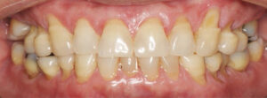
Noncarious cervical lesions (NCCLs) are defined as a loss of hard dental tissue near the cementoenamel junction, usually on the buccal surfaces of teeth, resulting in a grooved or wedge-shaped area of missing tooth structure. These lesions are increasing in prevalence, especially among adolescents and older adults.1 In particular, lifestyle changes — including the increased consumption of acidic drinks among younger patients and the number of older adults who take prescription medications, which can cause hyposalivation and xerostomia — raise the risk of NCCLs. Hyposalivation increases acidity in the oral cavity, softening the tooth surface and facilitating tooth loss (Figure 1).
A study reported that the prevalence of NCCLs in teenagers and adults ranges from 11% to 62%, and the number of lesions increases with age.2 Lesions most often occur on the buccal of maxillary premolars, but are also commonly found on other maxillary and mandibular anterior and premolar teeth. The exact etiology of NCCLs is unknown, but it is generally accepted that their cause is multifactorial.1,3,4 The clinical endpoint of NCCLs is the wearing away of dentition. The mechanisms involved include acid erosion (the loss of tooth structure by acid demineralization without the involvement of bacteria); abrasion (friction resulting in loss of tooth structure from dentifrices and toothbrushing); and abfraction (breaking away of tooth structure due to tensile stresses in the cervical area).1,3,4
Acid erosion is the most important factor involved in the development of NCCLs. Saliva normally keeps the oral environment in the 7.0 pH range (neutral) due to its ability to buffer acids. The source of acids can be intrinsic — bulimia or gastroesophageal reflux disease (GERD), for example — or extrinsic, such as acids from fruit juices, sports and energy drinks, and wine, to name a few. When oral acids bring the pH of the tooth surface below 4.5, demineralization of enamel and dentin can begin.5
EROSIVE CONCERNS
The action of strong acids causes the loss of calcium (inorganic matter) from the tooth surface, followed by a softening of the top layers of dentin and enamel. This softened layer then becomes susceptible to abrasive forces in the oral cavity and is easily removed, potentially causing wedge-shaped cervical lesions. The thin, softened layer is between 0.2 µm and 3 µm thick and is repeatedly removed by abrasive forces, leading to permanent loss of tooth volume in the cervical area.6
The pH (less than 4.5), duration and the strength (buffering capacity) of the acid challenge need to be considered. For example, cola (phosphoric acid) and certain energy drinks (citric acid) have a similar pH, but drinks containing citric acid are more erosive because more buffering is required to return the pH above the “critical” erosive point of pH 4.5.3 Regular and diet sodas and sports and energy drinks range from a pH of 2.5 to 3.4, respectively.7 Lemon, grapefruit and orange juices also cause erosive damage to enamel. Gastric acids from GERD are the most erosive, with a pH of 1.0 to 2.0.1 In addition, patients with gastric reflux often suffer from hyposalivation.8
Saliva plays an important role in oral health by cleansing the oral cavity and diluting and buffering acids. Individuals with xerostomia are much more susceptible to acid erosion. Patients are often advised to avoid brushing for 30 minutes to 60 minutes immediately after an acid exposure, presumably to allow softened surface enamel to remineralize — but, in truth, more effective preventive strategies (such as various fluoride-based therapies) should be recommended. The reality is that softened enamel is not remineralized by saliva over short time periods, and will be worn away even in the absence of tooth brushing by normal abrasive forces in the oral cavity, such as chewing and tongue action.
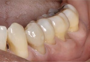
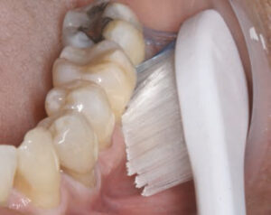
Dental abrasion, another contributing factor to NCCLs, is defined as wearing away of dental hard tissues by frictional forces.1 This can include wear caused by toothbrushing, flossing, tongue action, abrasive foods, and rubbing from opposing surfaces that are hard or rough, such as unpolished porcelain crowns.3 The effects of such actions are usually more pronounced on dentin surfaces than on enamel (Figure 2 and Figure 3).9
It appears that toothbrushing alone — regardless of the brush stiffness, filament size, aggressiveness or the frequency of brushing — causes little abrasive tooth wear.1 Rather, it is the abrasiveness of the toothpaste combined with brushing that is responsible for the loss of tooth structure.10,11 Dentifrices that include whitening agents or products promoted for their ability to control tartar tend to be more abrasive than other types. That said, all popular brands marketed in the U.S. today comply with abrasivity “safe limits” set by American Dental Association and International Organization for Standardization.12,13 Any toothpaste with a Relative Dentin Abrasivity (RDA) value of 250 or below can be used safely on a daily basis for a lifetime. Toothbrushes with stiffer bristles, however, have been linked to gingival recession.14
Abfraction is the microstructural loss of tooth structure due to excessive occlusal forces transmitted to the cervical area as flexural forces.1 These flexural forces, usually caused by cyclic loading, may lead to the breaking away of enamel rods in the cervical region of the tooth, causing microfractures of cementum and dentin.4 Bruxism may also contribute, although in the absence of acid there is minimal erosive tooth wear.15
Another possible modifying factor in abfraction formation involves the mobility of teeth and the effects of occlusal stresses in the formation of cervical lesions. A relationship has been found between tooth cervical lesions, tooth stability and periodontal support.15 Thus, as tooth mobility increases, teeth are actually less susceptible to excessive occlusal forces, as the teeth move under occlusal loads, resulting in less concentration of forces in the cervical areas.16
While these interocclusal relationships may be contributing factors in the formation of some NCCLs, it is likely that clinical signs of abfraction in cervical areas are not simply related to stress concentrations. Rather, they may play a role, along with acid erosion and toothbrush abrasion, in the multifactorial etiology of NCCLs.1,4
LESION MANAGEMENT
It’s important to note that NCCLs can occur at any time and may worsen throughout a patient’s lifetime. Without intervention, they can result in tooth fracture, hypersensitivity and cosmetic problems. Therefore, early diagnosis is important in order to initiate behavioral changes and avoid future invasive and expensive restorative interventions.
Clinical management of NCCLs includes first identifying the various etiologic factors present in a given patient. For example, if a patient is an active bruxer, has wear facets on the teeth, and presents with NCCLs, the clinician may decide to prescribe an occlusal splint or night guard.4 Occasionally, judicious occlusal adjustments of interferences may be performed. Acid intake must also be reduced.
Through the process of taking a comprehensive medical history, clinicians should question patients regarding their intake of acidic foods or beverages, including fruits, wine, and sports and energy drinks. It is important to monitor the frequency, duration, sequencing, and the mode of intake of these beverages. Sipping and swishing a drink, for example, are much more erosive than gulping the beverage, as the acidic beverage stays in contact with the tooth surface for longer periods.17 Consuming dairy after an acid challenge will help raise the pH and shorten the duration of erosive insult.18 Medication use, especially among older patients, should be reviewed, as drug-related xerostomia can contribute to tooth surface loss.19 Finally, patients should be asked about GERD, because there is a correlation between this disorder and dental erosion.20 These medical and behavioral factors have the net, and possibly cumulative, effect of demineralizing the tooth surface, which ultimately leads to the irreversible loss of surface enamel via abrasive forces such as toothbrushing, eating hard foods, and even cleaning with toothpicks. If the risk factors cannot be eliminated through behavioral changes or medical treatment, other preventive actions should be taken.
key takeaways
- Clinicians are seeing an increasing prevalence of noncarious cervical lesions (NCCLs), especially among adolescents and older adults.1
- Lesions most often occur on the buccal of maxillary premolars, but are also commonly found on other maxillary and mandibular anterior and premolar teeth.
- While the exact etiology of NCCLs is unknown, it is generally accepted their cause is multifactorial.1,3,4
- Early diagnosis is important in order to initiate behavioral changes and preventive therapies that can help patients avoid invasive restorative interventions.
- Treatment should include minimizing the intrinsic or extrinsic acids that are the primary cause(s) of the lesions.
- Brushing with a stannous fluoride toothpaste is a key element in prevention management.
PREVENTION AND TREATMENT
Conservative preventive measures using fluoride mouthrinses and topical fluoride should be professionally managed.21 The use of fluoride varnish and bonding agents can protect the teeth from acid attacks, but their long-term effects may be limited.22 Stannous fluoride is an important preventive tool to protect the tooth surface from an acid challenge.23,24 It provides superior protection for acid erosion versus sodium fluoride or monofluorophosphate products. It has been shown that a stabilized stannous fluoride toothpaste is effective in preventing erosive tooth wear.25 The mode of action of stannous fluoride is to form a thin layer of stannous-based complexes on the surface of enamel, protecting the surface from acid attack.26,27 The patient also receives the benefit of fluoride.26
Remineralization after an acid attack may be possible with the use of topically applied pastes containing casein phosphopeptides and amorphous calcium phosphate.28 These materials are supersaturated with calcium and phosphate and can “recharge” and remineralize demineralized tooth surfaces.
Treatment should include minimizing the intrinsic or extrinsic acids that are the primary cause(s) of lesions. Treatment for cervical hypersensitivity due to NCCLs may also be necessary. Using desensitizing dentifrices, such as those containing potassium nitrate or stannous fluoride, can be helpful.29 Other studies have shown that the use of a self-etching adhesive, sealant or resin-bonded flowable composite restoration may provide comprehensive and rapid control of hypersensitivity.30 More advanced lesions can usually be managed using resin-bonded composite restorations. In rare instances, a crown or porcelain veneer may be necessary.
Understanding the etiology of NCCLs and making a timely diagnosis will allow early and minimally invasive interventions, such as the use stannous fluoride toothpaste or fluoride varnish and sealants. Counseling for behavioral modifications — reducing the intake of acidic foods and beverages, for example — is of utmost importance. Emphasizing the need to take preventive measures to help avoid enamel erosion in the first place will help patients manage this common problem.
References
- Bartlett DW, Shah P. A critical review of non-carious cervical (wear) lesions and the role of abfraction, erosion and abrasion. J Dent Res. 2006;85:306–312.
- Huysmans MC, Chew H, Ellwood RP. Clinical studies of dental erosion and erosive wear. Caries Res. 2011;45(Suppl 1):60–68.
- Barbour ME, Rees GD. The role of erosion, abrasion and attrition in tooth wear. J Clin Dent. 2006;17:88–93.
- Grippo JO, Simring M, Coleman TA. Abfraction, abrasion, biocorrosion, and the enigma of noncarious cervical lesions: a 20-year perspective. J Esthet Restor Dent. 2012;24:10–23.
- Lussi A, Schlueter N, Rakhmatullina E, Ganss C. Dental erosion — an overview with emphasis on chemical and histopathological aspects. Caries Res. 2011;45(Suppl 1):2–12.
- Addy M, Shellis RP. Interaction between attrition, abrasion and erosion in tooth wear. Monogr Oral Sci. 2006;20:17–31.
- von Fraunhofer JA, Rogers MM. Effects of sports drinks and other beverages on dental enamel. Gen Dent. 2005;53:28–31.
- Campisi G, Lo Russo L, Di Liberto C, Di Nicola F, Butera D, Vigneri S, Comilato D, Lo Muzio L, Di Fedi O. Saliva variations in gastroesophageal reflux disease. J Dent. 2008;36:268–271.
- Lussi A, Schaffner M. Progression of and risk factors for dental erosion and wedge-shaped defects over a 6-year period. Caries Res. 2000;34:182–187.
- Moore C, Addy M. Wear of dentine in vitro by toothpaste abrasives and detergents alone and combined. J Clin Periodontol. 2005;12:1242–1246.
- Harpenau L, Noble W, Kao R. Diagnosis and management of dental wear. J Calif Dent Assoc. 2011;39:225–231.
- ISO Dentistry — Dentifrices — Requirements, test methods and marking. 11609:1995(e). Available at: http://www.iso.org/iso/iso_catalogue/catalogue_tc/catalogue_detail.htm? csnumber=38010. Accessed January 18, 2016.
- American Dental Association Acceptance Program Guidelines: Fluoride-Containing Dentifrices (2005). Available at: http://www.ada.org/~/media/ADA/Science%20and%20Research/Files/guide_fluoride_dentifrice.ashx. Accessed January 18, 2016.
- Niemi ML, Sandholm L, Ainamo J. Frequency of gingival lesions after standardized brushing as related to stiffness of toothbrush and abrasiveness of dentifrice. J Clin Periodontol. 1984;11:254–261.
- Brandini DA, Pedrini D, Panzarini SR, Benete IM, Trevisan CL. Clinical evaluation of the association of non-carious cervical lesions, parafunctional habits and TMD diagnosis. Quintessence Int. 2012;43:255–262.
- Kuroe T, Itoh H, Caputo AA, Nakahara H. Potential for load-induced cervical stress concentration as a function of periodontal support. J Esthet Dent. 1999;11:215–222.
- Grobler S, Jenkins G, Kotze D. The effects of the composition and method of drinking of soft drinks on plaque pH. Br Dent J. 1985;158:293–296.
- Lussi A, Jaeggi, T, Zero D. The role of diet in the etiology of dental erosion. Caries Res. 2004;38:34–44.
- Wiener RC, Wu B, Crout R, Wiener M, Plassmann B, Kao E, McNeil D. Hyposalivation and xerostomia in dentate older patients. J Am Dent Assoc. 2010;141:279–284.
- Farahmand F, Sabbaghian M, Ghodousi S, Seddighoraee N, Abbasi M. Gastroesophageal reflux disease and tooth erosion: a cross-sectional observational study. Gut Liver. 2013;7:278–281.
- Lussi A, Hellwig E. Risk assessment and causal preventive measures. Monogr Oral Sci. 2014;25:220–229.
- Austin RS, Stenhagen KS, Hove LH, et al. A qualitative and quantitative investigation into the effect of fluoride formulations on enamel erosion and erosion abrasion in vitro. J Dent. 2011;39:648–655.
- Lussi A, Carvalho TS. The future of fluorides and other protective agents in erosion prevention. Car Res. 2015;49(Suppl 1):18–29.
- Carvalho TS, Colon P, Ganss C, Huysmans MC, Lussi A, Schluete N, Schmalz G, Shellis RP, Tveit AB, Wiegard A. Consensus report of the European Federation of Conservative Dentistry: erosive tooth wear-diagnosis and management. Clin Oral Investig. 2015;39:277–283.
- Bellamy PG, Harris R, Date RF, et al. In situ clinical evaluation of a stabilized, stannous fluoride dentifrice. Int Dent J. 2014;64:43–50.
- Faller RV, Eversole SL. Protective effects of SnF2-Part III. Mechanism of barrier layer attachment. Int Dent J. 2014;64(Suppl 1):16–21.
- Algarni AA, Lippert F, Hara AT. Efficacy of stannous fluoride and their combination in dentin erosion prevention in vitro. Braz Oral Res. 2015;29:1–5.
- Cochrane NJ, Cai F, Huq NL, Burrow MF, Reynolds EC. New approaches to enhanced remineralization of enamel. J Dent Res. 2010;89:1187–1197.
- West NX, Seong J, Davies M. Management of dentine hypersensitivity: efficacy of professionally and self-administered agents. J Clin Periodontol. 2015;42(Suppl):S256–S302.
- Veitz-Keenan A, Barna JA, Strober B, et al. Treatments for hypersensitive noncarious cervical lesions: a Practitioners Engaged in Applied Research and Learning (PEARL) Network randomized clinical effectiveness study. J Am Dent Assoc. 2013;144:495–506.

More recent studies have pointed to a direct cause and effect relationship for NCCLs. That is the increasing incidence of sleep disordered breathing. Bruxism, especially during REM sleep, is conducted against tightly contracted masseters and internal pterygoid muscles. Lateral forces are significant and ultimately cause fracture of the enamel at the CEJ, producing the typical wedge-shaped loss of enamel. It is seen mainly in maxillary premolars and canines, but can be more generally spread directly proportional to the severity of bruxism and/ or sleep disrupted breathing.