
CAD/CAM Utility in Dentistry
Fadi AL Farawati, DDS, MS, MClinDent, and Edgar Davila, DDS, MS, CDT, FACP, share insights and perspectives from academia and private practice on dental applications for CAD/CAM technology in dental practice.
Fadi AL Farawati, DDS, MS, MClinDent, and Edgar Davila, DDS, MS, CDT, FACP, share insights and perspectives from academia and private practice on dental applications for CAD/CAM technology in dental practice
Whether a dental setting relies on laboratories or utilizes in-house equipment, digital scanning and computer aided design/computer aided manufacturing (CAD/CAM) is a staple of today’s practice. At the same time, rapid advances and open-architecture formats are increasing the utility of digital design and manufacturing technologies.
Clinicians have many options for the use of digital dentistry, ranging from restorative treatment and orthodontics to dental implants and oral surgery, to name a few. They also have flexibility in gradually implementing these technologies into practice. To help provide perspective, we asked two experts to share their insights.
Fadi AL Farawati, DDS, MS, MClinDent, of The Dental College of Georgia at Augusta University provides a practical and well-rounded academic view of the field, while Edgar Davila, DDS, MS, CDT, FACP, shares his acumen as a maxillofacial prosthodontist in private practice in Tampa, Florida, where he also maintains a full-service, commercial digital laboratory.
What is involved in terms of the equipment and training needed to incorporate digital scanning into daily practice? How would you describe the learning curve?
Fadi AL Farawati, DDS, MS, MClinDent: Clinicians can gradually incorporate digital scanning into practice, starting with leasing or financing a scanner and outsourcing production of dental prostheses to laboratories. Dentists also have the opportunity to purchase design software that will allow control over the final product design, which they can send to labs or produce themselves with in-house milling units or three-dimensional (3D) printers.
Digital dentistry requires a learning curve, of course, but manufacturers provide training and typically offer a supporting platform to resolve technical difficulties or design questions.
Edgar Davila, DDS, MS, CDT, FACP: The first step in incorporating digital scanning on a daily basis is the dentist must be convinced — based on existing scientific and clinical data — this is the best way to obtain intraoral impressions for producing predictable dentistry. Next, the clinician should partner with a manufacturer that will provide the intraoral scanner, along with training, support and service. The learning curve is faster as time progresses because the newer scanners are easier to use than earlier models. The scanning process is also faster and more comfortable for patients, too.
Compared to conventional techniques, what are the benefits, challenges and limitations of digital scanning?
FA: Accepting or rejecting digital scanning as a viable modality for a given office is determined by evaluating the time, financial and clinical advantages of the technology. These must be weighed against the classical barriers that face technology adapters, including:
- Time needed to learn and master the technology
- The investment in acquiring the equipment and, equally important, maintaining it. When considering long-term costs, be sure to calculate the cost of licensing/relicensing, updates, IT support, deprecation, security and patient privacy protection
- The ability and willingness to move beyond the clinician’s comfort zone
The digital scanning process has to be normalized and integrated fully in the practice to reap the maximum benefits. For example, this means all fixed-prostheses cases should be scanned, with traditional intraoral impressions taken on an as-needed basis. The benefits of scanning technology include:
- A positive impact on the patient experience
- Time savings and the ability to assess the scanned preparation and make corrections without redoing the impression
- Quality improvements realized through avoiding traditional accumulative errors while making a traditional impression and pouring the cast
- Financial considerations; more specifically, the elimination of conventional impression making, physical transportation of the impressions and casts, remaking costs, and associated labor. Digital scanning, by comparison, will positively impact practice revenue1
The main limitation to intraoral scanning is evident in removable prostheses, as the soft tissue capture is either overextended or underextended when scanners are used rather than traditional impressions after border molding. Acquiring an impression to fabricate a full-arch, implant-supported, fixed complete denture is more accurate with conventional impression techniques, as research shows less scanning accuracy when implants are diverted by 20 degrees or more.2
ED: In regard to tissue management, the techniques to obtain excellent impressions are the same for analog and digital approaches. Compared to conventional impressions, the benefits of digital scanning include efficiency, patient comfort, impression accuracy, and predictable restorations.
The challenges include the cost of the scanning system and how soon the dentist will apply it to most cases to recover the investment.
Limitations involve full-arch, fixed, splinted implant restorations, where scientific data show a conventional impression is more accurate. Another limitation lies in removable prosthodontics, where indirect scanning of a conventional impression with border molding produces the best clinical results.
Please briefly discuss various indications for CAD/CAM technology in dental practice.
FA: There are many, but here is a partial list:
- Manufacturing removable and fixed dental prostheses
- Fabricating orthodontic appliances, braces and various occlusal devices, such as occlusal guards and sleep apnea appliances
- Diagnosis by evaluating a digitally scanned area, or overlapping the scanned file with a digital radiograph
- Treatment planning by evaluating the available tooth structure, and soft and hard tissues through scanning and overlapping digital files
- A teaching aid at dental schools that’s used to evaluate, grade and self-assess student projects
ED: Specialized CAD software offers control of the final restoration design; this lets dentists create the proper morphology, cross-section, contours, occlusion and proximal contacts. Many dental procedures can be performed with CAD/CAM technology, including onlays, inlays, crowns, bridges, posts, implant crowns and bridges, implant bars, implant full-arch hybrids, implant full-arch ceramic bridges, occlusal splints, sleep apnea appliances, orthodontic retainers and aligners, complete dentures, partial denture frameworks, and implant surgical guides. This technology also facilitates implant treatment planning.
Office-based CAM systems are indicated when clinicians want to control the fabrication and finishing of the final restoration in-house. This allows same-day dentistry and faster delivery of final restorations. From single crowns to six-unit bridges, prostheses can be manufactured in the office with a three-axis or five-axis milling machine. Occlusal splints, working and study models, implant surgical guides, and temporary crowns and bridges can be either milled or 3D-printed in-house. In addition, orthodontic retainers and aligners can be fabricated over printed models.
Digital technology allows clinicians to work closely with labs when creating restorations, denture frameworks, implant abutments and similar items. What is the dentist’s role in terms of design input — and how does this process work?
FA: Dentists are encouraged to be part of the design process, as they are experts in the clinical situation and longevity of the produced product.
This process can be achieved with software that lets dental teams design the proposed final product and send the file to a lab or an in-house 3D printer or milling machine for fabrication. Alternatively, the lab can propose a design and send a file that can be viewed on a specific platform or program. The dentist approves the proposal or modifies it to produce the desired prosthesis.
ED: With crowns and bridges, the design of the morphology of the restoration is based on adjacent dentition and occlusion. Extensive anterior esthetic restorations or full-arch reconstruction (with or without changing the vertical dimension of occlusion) will require a diagnostic wax-up that will drive the final design of the restoration — and this is where dentists must work closely with the lab.
Denture framework fabrication requires the dentist to design the appliance, either digitally or in analog fashion, which will be transferred digitally by the lab technician before ultimately being approved or modified by the dentist.
Once given instructions, lab technicians can handle implant abutment and implant restoration design before sending it to the clinician for review and final approval.
What are the options for dentists to create their own designs in-house?
FA: Most manufacturers offer design software to dental teams. This will require training, of course, and it follows a learning curve, but this in-house approach provides better control of a final — and predictable — product. It is worth noting many of these programs require a financial commitment and relicensing. There are also different modules of these programs; depending on the office’s needs, acquiring a higher-level program might not be justified financially at the practice level.
ED: Multiple single crowns, onlays, inlays, single implants, screw-retained restorations over titanium-base abutments, and three bridges can be easily designed and manufactured in-house for offices with milling capabilities. These restorations can be stained and glazed at the practice for nonchallenging cases in which tooth shades are easily matched.
As noted, besides using labs, offices can utilize chairside milling units and 3D printers. What are the practical considerations, benefits and challenges associated with bringing manufacturing in-house?
FA: Adopting in-house manufacturing allows same-day restorations, which will increase revenue and boost patient satisfaction. In addition to milling units, 3D printers are producing long-term, U.S. Food and Drug Administration-approved products that can be used for provisionals and definitive complete dentures. Once the infrastructure and training are in place, it also allows offices to print implant surgical guides, as well as dentures, custom trays, orthodontic and occlusal appliances, wax-ups and provisional restorations.
ED: Same-day dentistry is becoming a more acceptable form of treatment. In fact, many patients ask to have restorations placed the same day, which often stems from previous experiences in which a temporary crown was lost while waiting for the permanent crown from the lab. To apply this practically, a dental assistant or technician with laboratory skills must be trained to scan, design and mill the prosthesis. The main benefit is patient satisfaction and attracting new patients who value clinically efficient care.
Although much of the focus is on milling units, 3D printers can create occlusal splints, models, custom trays, implant surgical guides, try-in dentures, temporary crowns, and definitive complete dentures. Dental materials used for 3D printing are improving, and this modality may eventually replace milled manufacturing for permanent crowns.
Please address the economic and practice efficiencies of digital technologies.
FA: Digital technologies are boosting practice efficiencies at the diagnostic, treatment planning and production levels, where quick decisions and immediate revisions can improve treatment predictability. For example, Joda and Bragger1 report that compared to conventionally manufactured implant crowns, digitally fabricated, implant-supported, single-unit reconstructions offer an 18% savings over the entire clinical and laboratory treatment process.
ED: The initial cost of an intraoral scanner can be easily recovered when the technology is used to the fullest. Unless otherwise indicated, this means using digital scanning for all crowns, bridges and smaller implant restorations. Another consideration is that besides an efficient and timely workflow, in-house restorations may be more precise due to fewer lab-associated steps and the potential for lab-induced errors during processing.
Any final thoughts?
FA: It is not a matter of “if” the profession will transition to intraoral scanning and full adoption of digital dentistry. The technology is here and improving on a daily basis. The real questions are when this complete transfer will happen, and the extent to which dental schools and practicing dentists will fully embrace it.
ED: The digital revolution is changing the world, and dentistry is no exception. The bottom line is that digital technology provides an opportunity to provide more precise and efficient care.
REFERENCES
- Joda T, Bragger U. Digital vs. conventional implant prosthetic workflows: A cost/time analysis. Clin Oral Implants Res. 2015;26:1430–1435.
- Papaspyridakos P, Gallucci GO, Chen CJ, Hanssen S, Naert I, Vandenberghe B. Digital versus conventional implant impressions for edentulous patients: Accuracy outcomes. Clin Oral Implants Res. 2016;27:465–472.
The authors have no commercial conflicts of interest to disclose.
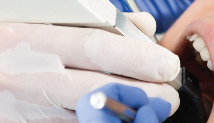
Featured image by MICROGEN/ISTOCK/GETTY IMAGES PLUS
From Decisions in Dentistry. March 2019;5(3):22–24.
