
 DANIELZGOMBIC/ISTOCK/GETTY IMAGES PLUS
10-Year Follow-Up on Resin Modified Glass Ionomer Restorations
This case study reports on the long-term clinical performance involving restoration with resin modified glass ionomer.
In 1972, a restorative material with unique and clinically useful features was created when researchers at the Laboratory of the Government in London, England, developed glass ionomer cement (GIC).1–4 This material was biocompatible and could develop stable, long-lasting chemical bonds to dentin. In addition, it did not require bonding agents and was not dependent on collagen and therefore not influenced by matrix metalloproteinases (MMPs).2,3,5–8 It exhibited resistance to caries through the uptake and release of fluoride, as well as its antibacterial properties.2,6,7,9–11 Clinical considerations for restoration techniques using GIC were described by McLean and Wilson in 1977.12
In 1988, a hybrid restorative material designated resin modified glass ionomer (RMGI) was developed by adding a small amount of resin to GIC.13 The resin allows for rapid setting induced by light curing, giving clinicians the ability to fully set the material in-office, thus preventing water absorption that can lead to problems with GICs.7 Chemically, RMGI adheres to dentin and its bond strength increases with time; it also exhibits higher flexural strength, and is less brittle and less sensitive to desiccation in the oral cavity than GIC.9,14
This paper will describe the long-term clinical results of restoration with RMGI in Class V preparations. At the time of the most recent follow-up, the patient’s restorations were more than 10 years old.

METHODS AND MATERIALS
In 2008, a 57-year-old male was seen in the emergency department for the extraction of two symptomatic teeth. Later, he became a patient at the University of the Pacific Arthur A. Dugoni School of Dentistry clinic in San Francisco, where he received a complete oral evaluation and treatment plan involving periodontal scaling and root planning, to be followed by the extraction of 10 nonrestorable teeth. The remaining teeth were #27, 26, 25, 24, 23, 22, 21 and 17.
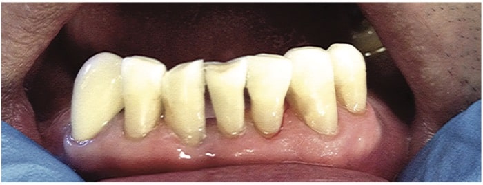
The scope of the treatment plan was determined by the restorability of the badly broken-down six lower anterior teeth (#27, 26, 25, 24, 23 and 22). Evaluation of these teeth revealed extensive loss of tooth structure, including compromised enamel to which composite resin could not successfully bond (Figure 1).
The lack of healthy enamel was cause for concern when considering restoration with resin composite. All teeth had a positive response to vitality testing. It was decided to restore teeth #26 to 21 with RMGI due to its ability to chemically bond to both dentin and enamel.2,9,15 Tooth 27 would be restored with a porcelain-fused-to-metal (PFM) crown.
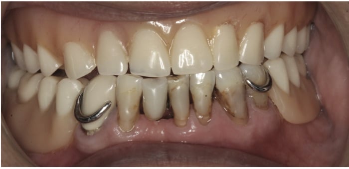
The task of restoring the lower anterior teeth was given to a senior dental student. Informed consent was obtained for restoration of the cervical lesions on teeth #26 to 21, as well as for the PFM crown and a removable prosthesis. Restoration with RMGI was completed in one clinic session on teeth #26 to 21. The cervical lesions were prepared using a carbide #4 round bur, ensuring that all of the dentin and peripheral enamel surfaces were instrumented. Next, they were cleaned and rinsed. Proceeding two teeth at a time, small, precut pieces of clear matrix were placed interproximally and secured with wedges. The prepped surfaces were again rinsed and dried without desiccating the exposed dentin. A conditioner was applied to the tooth surfaces following the manufacturer’s recommendations.16 The capsule of the restorative material was activated and the material was applied. The working time was adequate to fill, contour and adapt the material to the lesions in a controlled, well-paced manner. Once contoured, the RMGI was light cured for 20 seconds on each tooth. Using ultrafine diamond finishing burs under water spray and polishing strips, the restorations were polished and finished to the desired effect.16 The procedure was repeated for the remainder of the teeth (Figure 2).
RESULTS
Considering the condition of the teeth as presented, the initial restorative result was beyond what was expected. An upper denture and lower partial denture were later fabricated to replace the missing teeth. The lower anterior teeth were used not only to stabilize the lower partial, but also for their stability when chewing, thus subjecting them to strong flexural forces. The patient has been seen in the clinic on a regular schedule for periodontal and restoration maintenance, as well as follow-ups on the appliances.
On six of the recall appointments, the restored teeth were kept in good shape by bonding flowable resin composite or RMGI to the existing RMGI at the enamel margins.17,18 This was done to correct chipping where the incisal margin of the RMGI was bonded to the enamel. Caries were not involved.
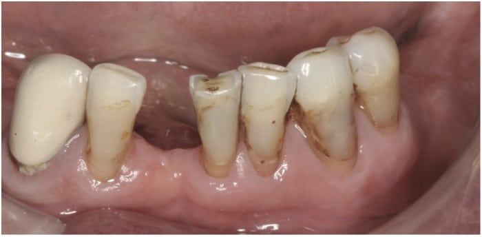
At five years post-op, tooth #25 was extracted due to periodontal disease. It was restored by modifying the existing lower partial denture. At the 10-year recall in January 2019, the five remaining heavily restored teeth still retained the bulk of the original RMGI that was placed in 2008. These teeth remained free of recurrent caries and continued to support a lower partial denture (Figure 3 and Figure 4). Although there was staining at the interface of the restored teeth and RMGI, the main concern at this juncture was controlling the patient’s periodontal disease.
DISCUSSION
The invention of GIC represented a new material with useful characteristics in restorative dentistry.1–4,9 This material was the result of an acid-base reaction involving polymers of polyacrylic acid and bases of fluoro-aluminosilicate.4 A chief benefit was the ability to avoid the problems associated with resin-dentin bonding involving collagen matrix and MMPs.5 Instead, GIC/RMGI are the only materials that attach chemically to tooth structure through bonds that get stronger over time.2,4,9 There are two interrelated methods of adhesion: micromechanical interlocking caused by glass ionomers being self-etching due to the polyacid component, and true chemical bonding that involves ionic bonds being formed between the carboxylate groups on the polyacid molecules and calcium ions in the tooth surface.
Over time, a diffusion process occurs between ions from the cement and tooth that move into an interfacial zone, creating an ion-exchange layer. The resulting structure causes the cement and tooth to adhere strongly.6 Through this process, adhesion of glass ionomer to tooth structure results in minimal microleakage.6,19 (The coefficient of thermal expansion of GIC and RMGI closely matches that of tooth structure, which explains their resistance to bacterial microleakage.) As noted, RMGI helps prevent caries through its fluoride release/recharge and antibacterial properties. It also has better flexural strength than GICs; by the same measure, it is hydrophilic, less soluble, and exhibits reduced surface crazing.2,9,14,20,21 A study of the amount of fluoride released by GIC versus RMGI did not find strong differences between the materials.22 The greatest fluoride release occurs during the first 24 hours, decreasing abruptly on the second day and tapering off until the seventh day.9,22
Unlike GIC, RMGI is a dual-set material that utilizes an acid-base reaction to set the glass ionomer component, while the resin component is set through light curing.4 The application of light enhances the resin’s initial set, thus reducing water loss during the chemical (acid-base reaction) phase. The setting time, however, is influenced by both reactions — which are, in a sense, competing with each other.4
It has been suggested that slightly delaying light activation — thus allowing the acid-base reaction to proceed (enhancing chemical bonding) — may result in a stronger set.4 As demonstrated in this report, RMGI can be a useful material when restoring carious and noncarious class V cases and other non-load-bearing lesions. In some situations, chemical bonding to tooth structure can alleviate or minimize the need for cavity preparation to achieve retention for restorative materials. The resin component in RMGI also allows for material repair with flowable resin composite and RMGI.17,18
RETENTION
Multiple studies have reported on the retention of RMGI used to restore cervical lesions. Due to concerns that such research is not calibrated as to the training and skills of the clinicians involved, citing studies on long-term outcomes can be problematic. For this reason, only a handful of studies are presented here.
A 5-year study by Smales and Ng23 reported median survival times of 30 months for one RMGI material and 42 months for another. Clinical research by Franco et al24 noted the clinical performance of RMGI was superior to resin composite restorations after five years. In a 6-year follow-up study, van Dijken25 found that RMGI showed an annual failure rate of 2%. A 7-year comparison study by Fagundes et al26 reported a retention rate of 30% for resin composite and 58% for RMGI. At the conclusion of a 13-year follow-up of six materials by van Dijken and Pallasen,27 RMGI posted a superior retention failure rate of 2.7%.
The long-term case report presented here demonstrates good results with RMGI restorations after 10 years. In the bigger picture, the initial goal of treatment was to gain time for the patient in a semi-comfortable situation with a partial denture. While a fixed solution would have been more desirable, it could not be realized for financial reasons. The repair of the lower anterior teeth and fully functioning partial denture have served the patient well. The decision to restore the anterior teeth has prevented massive bone loss that would have occurred had the teeth been extracted. As noted, since the initial restoration in 2008, the patient has lost one anterior tooth due to periodontal disease, and may lose more in the future. That said, the time gained in the transition process to a full lower denture contributed to the patient’s well-being and comfort. Proper eating and mastication have a major impact on patients and should be considered in the treatment planning process.
CONCLUSION
While RMGIs are often alluded to as inferior restorative materials because they are thought to be brittle and exhibit weak characteristics, they should be given consideration due to their unique advantages in specific circumstances. As shown in this 10-year case study, when treating teeth with advanced caries that are non-load bearing, susceptible to tooth flexure, have minimal enamel, and are subject to recurrent lesions, RMGIs may be an appropriate choice in restorative materials.
The authors have no commercial conflicts of interest to disclose.
Key Takeaways
- Following the introduction of glass ionomer cement (GIC) in 1972, resin modified glass ionomer (RMGI) was developed in 1988 by adding a small amount of resin to glass ionomer.
- The resin allows for light curing, giving clinicians the ability to fully set the material in-office, thus preventing water absorption that can lead to problems with GICs.7
- Chemically, RMGI adheres to dentin and its bond strength increases with time; it also exhibits higher flexural strength and is less brittle and less sensitive to desiccation in the oral cavity than GIC.9,14
- The coefficient of thermal expansion of GIC and RMGI closely matches that of tooth structure, which explains these materials’ resistance to bacterial microleakage.
- Both materials also help prevent caries through their fluoride release/recharge and antibacterial properties.22
- As demonstrated in this 10-year case study, when restoring teeth with advanced caries that are non-load bearing, susceptible to tooth flexure, have minimal enamel, and are subject to recurrent lesions, RMGI may be an appropriate choice in materials.
References
- Wilson AD, Kent BE. A new translucent cement for dentistry. The glass ionomer cement. British Dent J.1972;132:133–135.
- Ward D. The evolution of Glass Ionomer Restorative Materials. Available at: https://www.oralhealthgroup.com/features/evolution-glass-ionomer-restortive-materials/. Accessed September 9, 2020.
- Dentsply Chemfil Rock. Advanced Glass Ionomer Restorative. Available at: http://www.dentsply.se/bausteine.net/f/8746/SCChemFilRock110214.pdf?fd=2. Accessed September 9, 2020.
- Berzins DW, Abey S, Costache MC, Wilkie CA, Roberts HW. Resin-modified glass-ionomer setting reaction competition. J Dent Res. 2010;89:82–86.
- Osrio R, Yamauti M, Osorio E, Ruiz-Requena ME, Pashley D, Tay F, Toledano M. Effect of dentin etching and chlorhexidine application on metalloproteinase-mediated collagen degradation. Eur J Oral Sci. 2011;119:79–85.
- Sidhu SK, Nicholson JW. A review of glass-ionomer cements for clinical dentistry. J Funct Biomater. 2016;7:16.
- Croll TP, Berg JH. Glass-ionomer cement systems. Available at: https://www.aegisdentalnetwork.com/id/2010/09/glass-ionomer-cement-systems. Accessed September 9,2020.
- Margeas R. Simplified Delivery of a Resin-Modified Glass Ionomer Restorative. Available at: https://www.dentistrytoday.com/restorative/4727-simplified-delivery-of-a-resin-modified-glass-ionomer-restorative. Accessed September 9, 2020.
- Khoroushi M, Keshani F. A review of glass-ionomers: From conventional glass-ionomer to bioactive glass-ionomer. Dent Res J (Isfahan). 2013;10:411–420.
- Gao W, Smales R. Fluoride release/uptake of conventional and resin-modified glass ionomers, and compomers. J Dent. 2001;29:301–306.
- McComb D, Erickson RL, Maxymiw WG. A clinical comparison of glass ionomer, resin-modified glass ionomer and resin composite restorations in the treatment of cervical caries in xerostomic head and neck radiation patients. Oper Dent. 2002;27:430–437.
- McLean JW, Wilson AD. The clinical development of the glass-ionomer cement. II. Some clinical applications. Aust Dent J. 1977;22:120–127.
- Mitra SB. European Patent Application. Available at: https://patentimages.storage.googleapis.com/3f/a5/3b/5304673bb4f135/EP0323120A2.pdf. Accessed September 9, 2020.
- Mathis RS, Ferracane JL. Properties of a glass-ionomer/resin-composite hybrid material. Dent Mater. 1989;5:355–358.
- Burgess JO, Barghi N, Chan DC, Hummert T. A comparative study of three glass ionomer base materials. Am J Dent. 1993;6:137–141.
- GC America directions for using Fuji II LC Available at: www.gcamerica.com/products/operatory/GC_Fuji_II_LC_CAPSULES. Accessed September 9, 2020.
- Croll TP, Cavanaugh R. Resurfacing resin-modified glass-ionomer restorations. Inside Dent. 2009;5.
- Maneenut C, Sakoolnamarks R, Tvas M. The repair potential of resin-modified glass-ionomer cements. Dent Mater. 2010;26:659–665.
- Murray PE, About I, Franquin JC, Remusat M, Smith AJ. Restorative pulpal and repair responses. J Am Dent Assoc. 2001;132:482–491.
- Mitra SB. Adhesion to dentin and physical properties of a light-cured glass-ionomer liner/base. J Dent Res.1991;70:72–74.
- Uno S, Finger W, Fritz U. Long-term mechanical characteristics of resin-modified glass ionomer restorative materials. Dent Mater.1996;12:64–69.
- Cabral MF, Martinho RL, Guedes-Neto MV, Rebelo MA, Pontes DG, Cohen-Carneiro F. Do conventional glass ionomer cements release more fluoride than resin modified glass ionomer cements? Restor Dent Endod. 2015;40:209–215.
- Smales RJ, Ng KK. Longevity of a resin-modified glass ionomer cement and a polyacid-modified resin composite restoring non-carious cervical lesions in a general dental practice. Australian Dent J. 2004;4;196–200.
- Franco EB, Benetti AR, Ishikiriama SK, et al. 5-year clinical performance of resin composite versus resin modified glass ionomer restorative system in non-carious cervical lesions. Oper Dent. 2006;31:403–408.
- van Dijken JW. Retention of a resin-modified glass ionomer adhesive in non-carious cervical lesions. A 6-year follow-up. J Dent. 2005;33:541–547.
- Fagundes TC, Barata TE, Bresciani E, et al. Seven-year clinical performance of resin composite versus resin-modified glass ionomer restorations in noncarious cervical lesions. Oper Dent. 2014;39:578–587.
- van Dijken JW, Pallasen U. Long-term dentin retention of etch-and-rinse and self-etch adhesives and a resin-modified glass ionomer cement in non-carious cervical lesions. Dent Mater. 2008;24:915–922.
From Decisions in Dentistry. October 2020;6(9): 22-25.
