
CE Sponsored by Shofu—Clinical Update on Composite Restoratives
Choosing appropriate materials and using proper technique will help ensure successful outcomes in direct restoration procedures.
Choosing appropriate materials and using proper technique will help ensure successful outcomes in direct restoration procedures
PURCHASE COURSE
This course was published in the July 2017 issue and expires July 2020. The author has no commercial conflicts of interest to disclose. This 2 credit hour self-study activity is electronically mediated.
OBJECTIVES
After reading this course, the participant should be able to:
- Explain clinical management challenges and techniques when placing composite restorations.
- Describe the basic types of direct composite materials and considerations that affect the choice of materials for a given patient.
- Discuss the characteristics and ideal properties of composite resin materials, and advances in materials science.
INTRODUCTION
Informed patients seek treatments that are esthetic and have a beneficial influence on their overall health. To address the ever-growing needs of modern patients, Shofu has developed a score of award-winning dental products, including bioactive restorative materials, adhesive systems, minimally invasive abrasives, and a digital dental camera. These products are not only clinical problem-solvers, they also help practitioners expand the range of treatment techniques in a simple and cost-effective manner — elevating the practice’s growth and profitability. Giomer technology, a proprietary component of Shofu’s materials, combines the clinical benefits of bioactivity with superior physical, mechanical and esthetic properties. This innovative technology has been rigorously vetted in 13- and 8-year clinical studies that demonstrated intact esthetics and no issues with secondary caries, failures or postoperative sensitivity.*,**
REFERENCES
* Gordan VV, Mondragon E, Watson RE, Garvan C, Mjör IA. A clinical evaluation of self-etching primer and a giomer restorative material: results at eight years. J Am Dent Assoc. 2007;138:621–627.
** Gordan VV, Blaser PK, Watson RE, et al. A clinical evaluation of a giomer restorative system containing surface prereacted glass ionomer filler: results from a 13-year recall examination. J Am Dent Assoc. 2014;145:1036–1043.
Practical Considerations for Adhesive Dentistry
Dentists and materials scientists have long desired to find a less technique-sensitive and laborious approach to the placement of direct composite resin restorations. Unlike dental amalgam, which is a very forgiving material that allows simple placement, carving and finishing — with no intermediate bonding steps — placing composite resins is a more time-consuming process that requires additional adhesive steps, isolation from moisture contamination, and precise technique to achieve predictably successful long-term outcomes. Many of the earlier composite resins suffered from excessive polymerization shrinkage that further compromised the quality of the marginal seal and often led to postoperative sensitivity. Over the last few years, however, low-shrink and bulk-fill composites — including bioactive materials — have been developed that allow clinicians to more simply and efficiently place composite restorations and avoid some of these early pitfalls.
Compared to amalgam, placing composite requires more steps and exacting technique to achieve optimal results. Consequently, it has been suggested that resin restorations have a significantly higher risk of failure than amalgam restorations. Although some practitioners suggest the data does not agree with assumptions that composite is more difficult to work with, to a practicing dentist the challenges of placing successful composite restorations are obvious. The processes of adhesion, conditioning the tooth surface, and application of primers and adhesives — which is followed by the layering of composite resins — involves many steps that must be properly executed to ensure clinically acceptable results. For posterior composite restorations, in particular, the clinician must also manage challenges related to operator access and moisture control, as well as soft-tissue-related problems that often arise with Class II caries due to subgingival margin placement. Because composites are often placed in layers, numerous materials and steps are required to complete the procedure — all of which increase the potential for voids and marginal leakage that can ultimately lead to restoration failure.
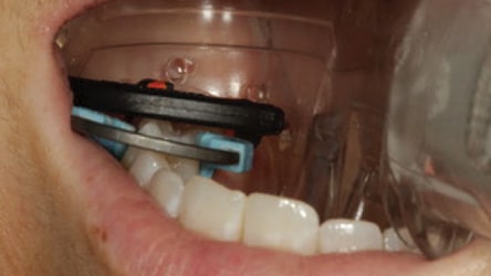
The complexity of the steps required to place composite contrasts sharply with those required for amalgam — although both materials require the use of a matrix system to restore anatomic proximal contours and contact areas, and prevent proximal gingival overhangs. For amalgam placement, a Class II procedure typically involves using a Tofflemire matrix band to contain the restorative material, which is then condensed into the preparation. Using an appropriate carving instrument, the restoration is carved into proper anatomic form. Next, occlusion is checked with articulating paper and the restoration is adjusted, as necessary. When placing composites, however, the clinician must first control the clinical environment using proper isolation techniques, such as a dental dam, isolation device or absorbent pad (Figure 1).
After isolating the operative area to control moisture contamination and using an appropriate matrix system (a sectional matrix, for example), each increment of composite placed typically requires: (1) etching, (2) rinsing, (3) drying, (4) desensitizing, (5) adhesive placement, (6) light curing, (7) placing a base or liner (if needed), (8) light curing, (9) placing the increment of composite, and (10) light curing. While use of single-bottle adhesive systems may eliminate some of these steps, even when using single-bottle adhesives it is generally agreed that etching the enamel using the selective-etch technique increases the efficacy of the resin bond to enamel. In addition, the antimicrobial agents found in desensitizers help inhibit bacterial growth and therefore can be considered useful when using self-etch, as well as total-etch bonding systems. Several increments of composites may be placed and cured before final sculpting of the occlusal surface of the restoration, checking and adjusting the bite, and placing an optional surface sealant to protect the marginal areas of the restoration.
GLASS IONOMER CEMENTS
Glass ionomer cement (GIC) and resin modified glass ionomer materials have been available for decades as dentin replacements and cavity liners, respectively, for use under direct restorative materials. With a coefficient of thermal expansion similar to dentin and the ability to recharge fluoride and remineralize tooth structure via ion exchange, glass ionomers have been the choice for many clinicians in deeper lesions where adhesive dentistry doesn’t fare as well. The “sandwich technique” utilizing GIC and composite resin was first described by McLean et al1 in 1985 and used GIC to bond the composite resin to teeth. Since that time, GIC has been widely used in large caries to replace lost dentin in “bulk” fashion.2–4
COMPOSITE CHARACTERISTICS
Amalgam is a material that can truly be condensed; in other words, when placing amalgam, the act of condensation actually pushes the alloy particles closer together. Condensing amalgam against a matrix band can actually cause deformation of the matrix, helping to create a contact with the adjacent tooth surface. In contrast, composite — as a class of materials — cannot be condensed. The material is pushed around the preparation with the placement instrument, but it is not condensed in any way. Short of using an additional instrument to physically push the composite and matrix against the adjacent tooth surface — and curing that increment immediately — proximal contacts cannot be consistently made by merely “pushing” the material into the cavity preparation in a fashion similar to placing amalgam. This makes contact placement and cavosurface marginal adaptation challenging.
Unfortunately, many dentists treat composite as if it were amalgam, and this is where problems arise. Because the viscosity of composites varies widely, clinicians can choose from a variety of materials based on operator preference. Heavy bodied or sculptable composites are generally chosen for posterior use, whereas creamier types are generally preferred for anterior esthetic composite work. Thanks to advances in materials science and efforts to find a truly universal material suited to both anterior and posterior applications, composite resins come in many forms, including flowables, packables, hybrids, microhybrids, nanomicrohybrids and microfills. In addition, new chemistries are being introduced as an alternative to the traditional bisphenol A-glycidyl methacrylate-based composites, reducing the problems of shrinkage, sensitivity and strength when used in posterior applications.
CLINICAL CHALLENGES
Establishing tight proximal contacts and eliminating voids between layers — while simultaneously seeking improving predictability and time efficiency in placement — represent significant challenges when placing composite restorations.
Voids: Recurrent decay, the leading cause of composite failure, is often traced to voids in the floor of the proximal box at the cavosurface margin of a Class II cavity,5 which is the class most vulnerable to incomplete filling. Although marginal adaptation can be verified visually, the incremental placement process — in addition to etching and bonding between layers — relies largely on blind faith that the area has been completely filled. These areas often cannot be seen radiographically unless placed in the same directional plane as the actual X-ray beam.
The traditional method of placing composite in increments no more than 2-mm deep was meant to facilitate curing and minimize polymerization shrinkage. The technique of layering, however, increases the potential to introduce voids into the restoration. Bulk-fill flowable composites are designed to serve as “dentin replacements.” In most cases, they can be utilized in increments up to 4 mm, although some materials are reportedly indicated for even deeper bulk placement. While bulk-fill flowables have the potential to minimize or eliminate voids by decreasing the number of layers in the restoration, typically they do not have the physical properties to withstand forces of occlusion, and therefore an additional capping layer may be required. This underscores the need to follow manufacturer instructions.
Replicating Proximal Contact and Contour: In most instances, the Tofflemire universal matrix commonly used for amalgam is not appropriate for use with composites. Clinicians can choose from a variety of sectional matrices designed specifically to replicate not just the proximal contact, but also the proximal contour. They are positioned between the restoring surface and adjacent proximal surface and use a ring to gently push the teeth apart, creating proximal contact and allowing for ideal anatomic placement of a sectional band. Concave on the inside and convex on the outside, the band can replicate the convex contour of the natural proximal tooth surface. It is the author’s experience that when a sectional matrix is placed correctly, less finishing with rotary instruments is needed because the intimate adaptation of the matrix to the cavity preparation walls creates less chance of composite excess or overhangs.
Depth of Cure: According to a study by Campodonico et al,6 “When using resin-based composite restorative materials, clinicians should be more concerned about the effect of filling techniques on curing depth than about how these techniques affect shrinkage stress.”6
INCREMENTAL LAYERING VERSUS BULK FILL
As noted, clinical placement of composites traditionally has been accomplished using an incremental placement technique. Due to polymerization shrinkage — as well as the inability to light cure composite materials beyond a certain depth — clinicians were generally advised to place composite resin in increments of 2 mm or less. Yet given advances in polymer chemistry, photo activation and curing-light technologies that we see with today’s composite systems, is this still true? Studies of incremental versus bulk-fill placement of composite show there is no difference in cuspal deflection or marginal integrity when comparing placement techniques. The main clinical issue with bulk-fill materials is depth of cure. It is also important to note that directional curing from the buccal and lingual (palatal) aspects after removal of the matrix help increase clinicians’ ability to cure composite at the gingival margin of the proximal box in a Class II restoration.7–9
BULK-FILL FLOWABLES AS DENTIN REPLACEMENTS
Bulk-fill flowable composites were introduced in 2011 and are indicated for use as a bulk-fill dentin replacement in posterior restorations. The ability to place bulk fills in increments of up to 4 mm (or greater, depending on the material) is a significant time-saver because it reduces the number of potential layers of the standard 2-mm increment thickness in half. While this concept sounds simple, there are several important factors a material must meet in order to warrant this type of indication. Ideally, bulk-fill composites should offer increased depth of cure, a viscosity that readily adapts to the internal geometry of the cavity preparation (without the need for extra manipulation of the material), and low rates of polymerization shrinkage and shrinkage stress.
Because of their opacity and percentage of fillers, bulk-fill flowable composites may require a conventional composite material to be placed as the final (i.e., occlusal) increment.10–13 As previously noted, however, it is important to consult manufacturer instructions for all materials used.
‘SMART’ COMPOSITES
Marginal breakdown and recurrent decay are two processes by which restorations can ultimately fail. One of the challenges in developing restorative materials is to find a mechanism by which the materials themselves can slow or interfere with the attraction of biofilm to tooth structure, and/or buffer the effects of acid metabolism. Glass ionomers work by releasing fluoride ions to help make tooth structure more resistant to acid attack. Because of the solubility of the material in the oral environment, there is often a short period that fluoride protection can be available until the material “recharges” via external sources, such as fluoride dentifrice. One group of materials utilizes proprietary Giomer chemistry that features a surface pre-reacted glass filler within the composite resin matrix. With this technology, molecules of polyacrylic acid solution permeating through the surface-modified layer reach the multifunctional fluoro-boro-alumino-silicate glass core and form a trilaminar structure that facilities the release and recharge of fluoride, strontium, borate, aluminium, silicate and sodium (natrium). Ionic exchange observed in the Giomer may help buffer or neutralize acid byproducts of bacterial metabolism for extended periods over the life of restoration. This makes Giomer materials especially effective for use in caries-prone individuals, who often exhibit extreme periods of acid challenge to unrestored teeth and the marginal interfaces of restored teeth.14–17
NANOHYBRID COMPOSITE TECHNOLOGIES
Nanohybrid and nanofilled composites are now common and are more highly filled and polishable than previous generations of materials. These products can be used in the posterior region, as well as in anterior esthetic areas. Many contain nanofiller technology and are formulated with nanomer- and nanocluster-filler particles. Nanomers are separate, discrete nanoagglomerated particles of 20 to 75 nanometers in size, while nanoclusters are loosely bound agglomerates of nanosized particles. The combination of nanosized particles and nanocluster formations reduces the spacing of the filler particles, thus creating the ability to increase filler capacity while still offering excellent polishability. Formulated for increased luster and polish retention,18–20 nanohybrids have physical properties that are superior to earlier generations of microhybrid composites.
BULK-FILL COMPOSITE RESINS
As noted, bulk-fill composites help simplify placement by decreasing the number of incremental layers that must be placed and individually cured. In order to facilitate successul outcomes in bulk-fill procedures, these materials must be formulated to allow for increased depth of cure while having less shrinkage and shrinkage stress than previous generations of composites. One way this has been accomplished is by increasing the amount of (or by using different) photoinitiators to allow for increased depth of cure. In addition, other types of monomers and elastic fillers have been developed that minimize shrinkage and shrinkage stress when the material is polymerized. One result of providing a deeper depth of cure, however, is often having to settle for a more translucent, less “chameleon-like” quality to the material. In other words, a key challenge in formulating a bulk-fill materials is achieving an opacity that retains the esthetic quality of the material, but without compromising depth of cure.21,22
LOW-SHRINK COMPOSITES
Polymerization shrinkage of composite resins has been a topic of discussion for many years. Early failures of composite materials have been attributed to the shrinkage of the resin during the curing phase, which opens a gap between the composite and tooth where bacterial access could lead to microleakage and recurrent decay. A goal of material science has been to create materials using polymers that do not shorten or shrink during photopolymerization. Some of the clinical problems associated with polymerization shrinkage stress in composite resins include:
- Cracks in teeth and deformation of cusps
- Formation of “white lines” (i.e., gaps) between the restorative material and preparation margin
- Cuspal deflection, leading to postoperative sensitivity under occlusal load
- Debonding of restorations.
These phenomena can also lead to marginal leakage, secondary caries and, ultimately, restoration failure.
The amount a composite will shrink varies and depends on the monomer used, the type and amount of fillers, and how the fillers and resin matrix interact. Manufacturing modifications to reduce polymerization shrinkage may include: increasing the filler load, while decreasing the amount of monomer; incorporating prepolymerized fillers, which maintain their volumetric dimension, thereby reducing shrinkage; and using monomers of higher molecular weights, which shrink less. Recognizing the clinical challenge posed by shrinkage, material manufacturers have answered by producing low-shrink composites that help minimize the potential negative effects of polymerization-induced shrinkage stress.
At the same time, manufacturers are working to produce materials that offer superior wear resistance, color stability, easy polishability and high gloss retention. Specific formulations may provide additional clinical advantages; for example, a low-shrink composite has recently been introduced that offers Giomer technology, adding a biologic benefit that helps resist acid attack at the tooth/restorative interface — particularly in patients at high caries risk.
PLACEMENT TECHNIQUE FOR A LOW-SHRINK, CLASS II COMPOSITE RESTORATION
A layering method recommended by the author to eliminate voids in difficult-to-access gingival portions of a Class II caries preparation starts with the use of a flowable composite on the gingival floor of the proximal box, and at the angles where the gingival floor meet the facial and lingual vertical walls. This is the area where most layered composites will have voids and ultimately fail from recurrent decay. Using a fluoride-releasing flowable in this area may offer increased protection due to its ability to inhibit plaque accumulation.
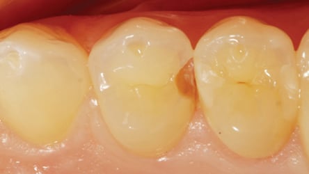
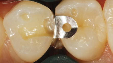

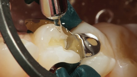
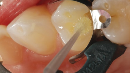
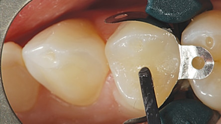
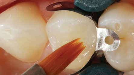
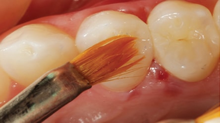
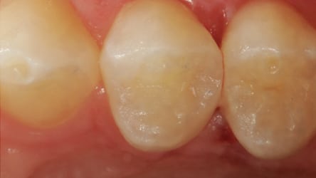
Figure 2 shows an occlusal view of tooth #12 that had a previous composite restoration that had debonded and was lost. After isolation, the tooth was prepared for a Class II composite restoration and a sectional matrix, cervical wedge and ring were applied to the distal aspect (Figure 3). A selective etch of the enamel was made using 37% phosphoric acid for 15 seconds, before rinsing with water (Figure 4 and Figure 5). The bonding resin was applied (Figure 6) with a microbrush applicator. Once the application was complete, the bonding resin was light cured for 30 seconds (Figure 7). Next, a low-shrink composite was placed (Figure 8) and light cured in successive 2-mm increments, filling the remainder of the proximal box and occlusal portion of the preparation. The final increment was sculpted into the proper anatomic form and brushed lightly with a sable brush to smooth the composite prior to light curing (Figure 9).

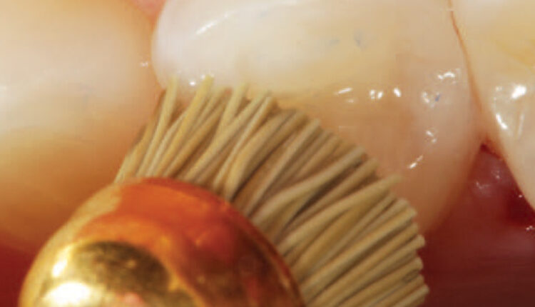
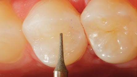
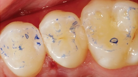
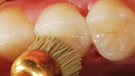
Rotary composite finishing instruments (such as elliptical or ovoid shapes that can impart a concavity) will ruin the anatomical form and are not recommended. An interproximal finishing instrument, such as a “needle” or “mosquito” diamond, is an excellent choice for use on the occlusal surface for minor adjustment, as it will impart a natural convexity to the cusp as the adjustment is made (Figure 10). Occlusion should be checked with articulating paper and any necessary adjustments made without ablating the anatomy of the restoration (Figure 11). Once the final occlusal contour is achieved, finishing and polishing can be completed using rubber abrasives and polishing brushes (Figure 12) that reach the nuances of the occlusal surface and marginal ridges. The surface of the restoration should be re-etched for a few seconds to ensure cleanliness, followed by the application of surface sealant (Figure 13) to fill any microscopic discrepancies that can’t be seen, but may exist. Finally, the sealant is air thinned and light cured from occlusal and proximal aspects. Figure 14 shows an occlusal view of the completed, Class II low-shrink composite restoration.
CONCLUSION
The ultimate goal in the creation of composite materials is to simplify the placement of direct composite resins without compromising the quality of the final result. Trying to recreate natural tooth structure with manmade materials is no easy task. Yet, advances are being made that allow clinicians to deliver esthetic restorations using tooth-colored restorative materials that offer patients a functionally esthetic, long-lasting result. Materials science continues to develop and refine resin materials that require fewer steps to place, are less technique sensitive, are more biologically harmonious with natural teeth, and are “smart” in the way they can help protect teeth and even restorations themselves.23
REFERENCES
- McLean JW, Powis DR, Prosser HJ, Wilson AD. The use of glass-ionomer cements in bonding composite resins to dentine. Br Dent J. 1985;158:410–414.
- Mount GJ. Clinical placement of modern glass ionomer cements, Quintessence Int. 1993;24:99–107.
- Christensen G. Glass-ionomer-resin restorations. Clin Res Assoc Newsletter. 1992;16(3):1–2.
- Mount GJ. Clinical requirements for a successful “sandwich” — Dentine to glass ionomer cement to composite resin. Aust Dent J. 1989:34:259– 265.
- Bouillaguet S, Ciucchi B, Jacoby T, Wataha JC, Pashley D. Bonding characteristics to dentin walls of class II cavities, in vitro. Dent Mater. 2001;17:316–321.
- Campodonico CE, Tantbirojan, D, Olin PS, Versluis A. Cuspal deflection and depth of cure in resin based composite restorations filled by using bulk, incremental, and trans tooth illumination techniques. J Am Dent Assoc. 2011;142:1176–1182.
- Rees JS, Jagger DC, Williams DR, Brown G, Duguid W. A reappraisal of the incremental packing technique for light cured composite resins. J Oral Rehab. 2004;31:81–84.
- Flury S, Hayoz S, Peutzfeldt A, Hüsler J, Lussi A. Depth of cure of resin composites: is the ISO 4049 method suitable for bulk fill materials? Dent Mater. 2012;28:521–528.
- El-Safty S, Silikas N, Akhtar R, Watts DC. Nanoindentation creep versus bulk compressive creep of dental resin-composites. Dent Mater. 2012;28:1171–1182.
- Ilie N, Bucuta S, Draenert M. Bulk-fill resin-based composites: an in vitro assessment of their mechanical performance. Oper Dent. 2013;38:618–625.
- Juloski J, Carrabba M, Aragoneses JM, Forner L, Vichi A, Ferrari M. Microleakage of Class II restorations and microtensile bond strength to dentin of low-shrinkage composites. Am J Dent. 2013;26:271–277.
- Van Ende A, De Munck J, Van Landuyt KL, Poitevin A, Peumans M, Van Meerbeek B. Bulk-filling of high C-factor posterior cavities: effect on adhesion to cavity-bottom dentin. Dent Mater. 2013;29:269–277.
- Roggendorf MJ, Krämer N, Appelt A, Naumann M, Frankenberger R. Marginal quality of flowable 4-mm base vs. conventionally layered resin composite. J Dent. 2011;39:643–647.
- Nakamura N, Yamada A, Iwamoto T, et al. Two-year clinical evaluation of flowable composite resin containing pre-reacted glass-ionomer. Pediatr Dent J. 2009;19:89–97.
- Tamura D, Saku S, Yamamoto K, Hotta M. Saliva protein which adsorbs to composite resin containing S-PRG filler. Japanese Soc Conservative Dent. 2010;53:191–206.
- Saku S, Kotake H, Scougall-Vilchis RJ, et al. Antibacterial activity of composite resin with glass-ionomer filler particles. Dent Mater J. 2010;29:193–198.
- Izono T, Saku S, Yamamoto K. Application to the tooth coating material of the glass filler containing acid reactive fluoride. Japanese Soc Conservative Dent. 2009;52:237–247.
- Rodrigues SA Jr, Ferracane JL, Della Bona. Flexural strength and Weibull analysis of a microhybrid and a nanofill composite evaluated by 3- and 4-point bending tests. Dent Mater. 2008;24:426–431.
- Curtis AR, Palin WM, Fleming GJ, Shortall AC, Marquis PM. The mechanical properties of nanofilled resin based composites: characterizing discrete filler particles and agglomerates using a micromanipulation technique. Dent Mater. 2009;25:180–187.
- Beun S, Glorieux T, Devaux J, Vreven J, Leloup G. Characterization of nanofilled compared to universal and microfilled composites. Dent Mater. 2007;23:51–59.
- Yapp R, Powers JM. Depth of cure of several composite restorative materials. Dental Advis Res Rpt. 2011;33(2):1.
- Alrahlah A, Silikas N, Watts DC. Post-cure depth of cure of bulk fill dental resin-composites. Dent Mater. 2014:30:149–154.
- Lowe RA. Advances in composite resin materials. Inside Dent. 2015;11(12):45–52.
From Decisions in Dentistry. July 2017;3(7):37–42.

