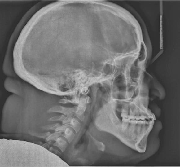
Return to "Understanding Orthognathic Surgery" FIGURE 6. Posttreatment cephalometric radiograph illustrating proclination of the maxillary incisors and uprighting of the mandibular incisors in the correction of Class III patient using orthodontics only. Next Previous