

A Review of Esthetic Crowns for the Primary Anterior Dentition
An exploration of esthetic crown options for deciduous teeth, and the evidence supporting their use.
An exploration of esthetic crown options for deciduous teeth, and the evidence supporting their use
In modern society, the demand for esthetic restorations is increasing in pediatric dentistry. Parents have high expectations of the restorative materials, especially in the esthetic zone. Durability, cost and esthetics are the main factors considered by parents when determining a treatment plan.1 Tooth-colored restorations, especially for anterior teeth, are of primary concern to parents. A particular study showed that parents ranked attractiveness and health similarly.2 The same study also mentioned that parents viewed silver stainless steel crowns as unhealthy and esthetically unacceptable. While the decision on treatment planning is often driven by the parents,3 children are also concerned about esthetics and tend to prefer white fillings rather than silver.4
Minimal research is available to provide evidence of the comparative clinical success of various esthetic crowns for primary teeth. Recommendations for esthetic full coverage crowns in the primary dentition are based on limited research, mainly relying on retrospective studies and case reports. Well-controlled randomized clinical trials are difficult to implement in children due to financial, behavioral and social barriers.
This article will discuss the possible esthetic crown options in anterior teeth, and the available evidence to support the use of various restorations.
STAINLESS STEEL CROWNS AND OPEN-FACED STAINLESS STEEL CROWNS
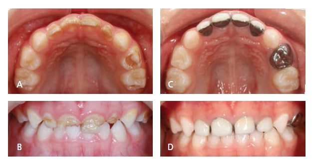
FIGURES 1A THROUGH 1D COURTESY YI-CHUN CHEN, DDS
Stainless steel crowns (SSCs) can restore anterior teeth with extensive caries but still provide a stable restoration. This type of crown is resistant to fracture and can be easily fitted as well as crimped on all surfaces, even when there is minimal remaining tooth present.5 The dissatisfying silver color can be improved by the open-face stainless steel crown technique.6,7 After the recommended glass ionomer cement is set, the metal on the facial surface of the crown is removed by a 330 bur. Tooth-colored resin is placed after etching and bonding (Figures 1A through 1D). This open-face technique is inexpensive and can provide a better esthetic appearance compared to the original silver metal color — however, it is time consuming, and esthetics are compromised by the metal margin. Hemorrhage control is extremely important when placing the resin, and it is often difficult to control bleeding after the SSC tooth preparation. SSCs are less frequently used in primary anterior teeth than posterior teeth, but due to the occlusion or extensive caries, they are sometimes used in anterior teeth. SSCs are more commonly applied to the mandibular incisors, where esthetics is less noticeable. The retention rate of SSCs is reported as 93% after 27 months and SSCs appear to be retained significantly longer than preveneered SSCs.8
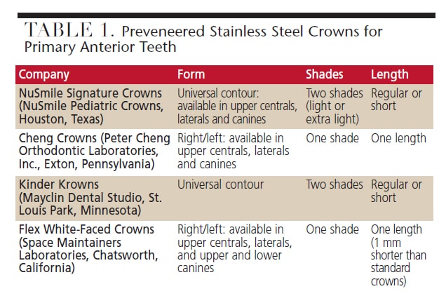
PREVENEERED STAINLESS STEEL CROWNS
Preveneered SSCs were introduced into the pediatric dentistry market to provide another esthetic option for anterior restorations. These crowns have prefabricated tooth-colored material bonded to SSCs on the facial surface, usually with resin-based composite material. Various brands are listed in Table 1. The prefabricated esthetic facial surface enables the crown to be placed in an area with poor hemorrhage control, providing a white appearance (Figures 2A and 2B).9–11
FIGURES 2A AND 2B COURTESY JEROD BRAZEAL, DDS
The main problem with these crowns is the possible wear on the incisal edge, as well as partial or total fracture of the facial portion. It is difficult to repair the chipped portion and if parents or children are concerned about a portion of metal showing, the replacement of the whole crown is needed. Several studies have shown that 12% to 39% of preveneered crowns have wear or partial facing loss.9–11 Total loss of the esthetic facing was reported to range up to 24% of the preveneered crowns in these studies.
Crimping of the facial preveneered portion will cause esthetic facing to fracture easily. Thus, retention of the crowns relies on crimping the lingual portion of the crown. When applying the crown, it must not be forced over the prepared tooth because the prefabricated resin portion is easily fractured. When cementing these crowns, it is recommended to use glass ionomer cement to hold the crown in place until the cement sets. The retention rate for this type of crown is reported to be above 90% after six to 17 months.8–11
A recent prospective study compared parental satisfaction of three different tooth-colored anterior crowns.12 The study showed that parents are least satisfied with preveneered SSCs when compared with zirconia and strip crowns, mainly due to the color. In another study by Champagne et al,13 however, the parental satisfaction was 93% at 13 months post crown placement. Even though the lingual portion of the crown was silver, parents had high satisfaction with these crowns.
STRIP CROWNS
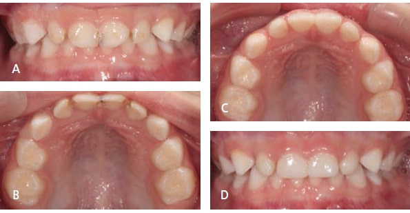
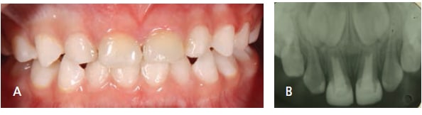
This type of crown is composed of celluloid crown forms that act as matrix forms to fill with tooth-colored materials. Usually, these crowns are restored with resin-based composite to allow for selection of shades to match the adjacent teeth and provide an excellent esthetic outcome (Figures 3A through 3D). Resin modified glass ionomer cement has been used for the material as an interim restoration in younger children.14,15 Placing a strip crown is technique-sensitive because moisture and hemorrhage control are very important to prevent contamination of the resin with blood or saliva. There must be enough remaining tooth structure to allow for adequate bonding.5 After the polymerization of the resin-based composite, the celluloid form can be easily removed with a dental bur or a sharp blade.
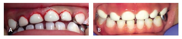
When placing strip crowns over pulpectomy treated teeth obturated with iodoform paste, it is important to use opaque resin or remove iodoform paste below the cervical area so the yellow iodoform paste will not be seen through the strip crown from the facial surface (Figures 4A and 4B).16 Likewise, teeth that have been restored with zinc-oxide eugenol after pulpal treatment should have a glass ionomer or resin modified glass ionomer base placed over the zinc-oxide eugenol so that polymerization upon light exposure is not compromised. Overall, parental satisfaction was excellent for this type of crown. One study showed that parents preferred strip crowns to preveneered crowns.12 However, partial or total loss of the crown or durability was the main concerns from parents.12,17 Studies show that the overall retention rate was above 80% after 18 to 24 months.16,18
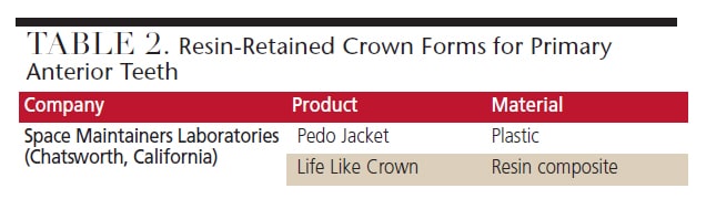
RESIN-RETAINED CROWN FORMS
Besides the widely used strip crowns, there are two alternatives that bind to the tooth structure (Table 2). One is made of a tooth-colored plastic. After the tooth is etched and bonded, a crown filled with resin material is fitted to the tooth. There is no need to remove the crown form after polymerization. However, the crown cannot be reshaped with a bur because the plastic material will disform from the heat. The other alternative is made of resin composite material and can be reshaped with a finishing bur to provide a more esthetically pleasing appearance. If the tooth is not reduced adequately, forcing the crown down to the tooth can cause the crown to crack or fracture.
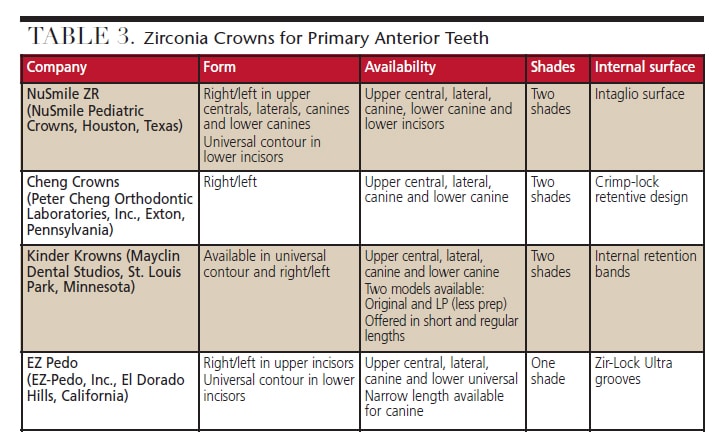
ZIRCONIA CROWNS
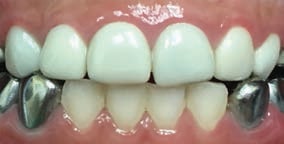
FIGURE 5 COURTESY MARIA JOSE CERVANTES MENDEZ, DDS, MS
Zirconia crowns have been successfully used for permanent teeth for many years and provide excellent esthetics due to their natural appearance. Zirconia crowns are relatively new in the practice of pediatric dentistry, introduced in 2010. They are recommended for both anterior and posterior cases. Table 3 lists the products available in this category for the primary anterior dentition. Zirconia crowns are very strong, durable and esthetically pleasing (Figure 5). Unfortunately, since these preformed crowns cannot be crimped, the retention is reliant on the internal surface designs (Table 3) and cementation.
There is a learning curve for dentists who are unfamiliar with placing pediatric zirconia restorations. It may take longer to prepare the tooth and fit the crown because the tooth is prepared to fit the crowns rather than simply adjusting crowns to fit a tooth. Zirconia crowns require a feathered margin as in other crown preparations; however, these crowns need more tooth reduction when compared to strip crowns and SSCs. One study showed that there is no significant difference in tooth reduction for anterior zirconia crowns between the four brands available.19
Hemorrhage control is important to prevent contamination of the crowns and achieve adequate bonding.20 Generally, these crowns require thorough cleaning with alcohol before final cementation, although one brand includes try-in crowns for trial fitting, which allows the final crown to remain contamination-free prior to cementation. Most manufacturers recommend glass ionomer cement or resin modified glass ionomer to cement zirconia crowns — however a bioactive resin cement has also been recommended by a manufacturer.
There are very few studies evaluating the clinical use of zirconia crowns in primary anterior and posterior teeth.12,21–24 One randomized controlled trial with a short follow-up period of six months was conducted.22 In this study, 129 teeth were randomly distributed into one of the three treatment groups (strip crowns, preveneered SSCs or zirconia crowns). Zirconia crowns showed the highest retention rate (100%) and better gingival health adaptation, whereas preveneered SSCs had a 95% retention rate and strip crowns had a retention rate of 78%.
A retrospective study was performed to examine 44 anterior crowns for an average of 20.8 months after placement.21 There was no wear observed on any teeth opposing the crowns. Among the parents/caregivers in the study, 78% reported that the crowns improved appearance. The overall satisfaction from parents/caregivers with these crowns was very high (9.3 out of possible 10).
A prospective study to compare parental satisfaction was conducted using three different tooth-colored anterior crowns.12 This study showed that parents had the highest satisfaction with zirconia crowns, followed by strip crowns and preveneered SSCs.
KEY TAKEAWAYS
- Stainless steel crowns are a stable restoration option for anterior teeth with extensive caries. The open-face technique can provide a better esthetic appearance compared to the original silver metal color.
- A prefabricated esthetic facial surface enables preveneered stainless steel crowns to be placed in an area with poor hemorrhage control, providing a white appearance. The main problem is the possible fracture of the facial portion, which is difficult to repair.
- Strip crowns are used in cases with enough remaining tooth structure to allow for adequate bonding. Placing strip crowns is technique-sensitive because moisture and hemorrhage control are important to prevent contamination of the resin with blood or saliva.
- Zirconia crowns are relatively new in pediatric dentistry and require a learning curve for dentists. These crowns cannot be crimped, and hemorrhage control is important to prevent contamination of the crowns and achieve adequate bonding.
CONCLUSION
Clinically, the choice of the crown depends on the preference of the operator, behavior of the patient, occlusion, amount of remaining tooth structure, and parental expectations. There has yet to be a clinical study to show which restoration is considered the best. Further well-controlled randomized clinical trials are needed to enable clinicians to make the best evidence-based decisions.
REFERENCES
- Tinanoff N, Douglass JM. Clinical decision-making for caries management in primary teeth. J Dent Educ. 2001;65:1133–1142.
- Woo D, Sheller B, Williams B, Mancl L, Grembowski D. Dentists’ and parents’ perceptions of health, esthetics, and treatment of maxillary primary incisors. Pediatr Dent. 2005;27:19–23.
- Zimmerman JA, Feigal RJ, Till MJ, Hodges JS. Parental attitudes on restorative materials as factors influencing current use in pediatric dentistry. Pediatr Dent. 2009;31:63–70.
- Fishman R, Guelmann M, Bimstein E. Children’s selection of posterior restorative materials. J Clin Pediatr Dent. 2006;31:1–4.
- Waggoner WF. Restorative dentistry for the primary dentition. In: Casamassimo PS, Fields H, McTigue D, Nowak A, eds. Pediatric Dentistry: Infancy Through Adolescence. 5th ed. Phiadelphia, Penn: WB Saunders Co/ Elsevier Inc; 2013:304–332.
- Hartmann CR. The open-face stainless steel crown: an esthetic technique. ASDC J Dent Child. 1983;50:31–33.
- Helpin ML. The open-face steel crown restoration in children. ASDC J Dent Child. 1983;50:34–38.
- Lopez-Loverich AM, Garcia MM, Donly KJ. Retrospective study of retention of stainless steel crowns and pre-veneered crowns on primary anterior teeth. Pediatr Dent. 2015;37:530–534.
- Roberts C, Lee JY, Wright JT. Clinical evaluation of and parental satisfaction with resin-faced stainless steel crowns. Pediatr Dent. 2001;23:28–31.
- Shah PV, Lee JY, Wright JT. Clinical success and parental satisfaction with anterior preveneered primary stainless steel crowns. Pediatr Dent. 2004;26:391–395.
- MacLean JK, Champagne CE, Waggoner WF, Ditmyer MM, Casamassimo P. Clinical outcomes for primary anterior teeth treated with preveneered stainless steel crowns. Pediatr Dent. 2007;29:377–381.
- Salami A, Walia T, Bashiri R. Comparison of parental satisfaction with three tooth-colored full-coronal restorations in primary maxillary incisors. J Clin Pediatr Dent. 2015;39:423–428.
- Champagne C, Waggoner W, Ditmyer M, Casamassimo PS, MacLean J. Parental satisfaction with preveneered stainless steel crowns for primary anterior teeth. Pediatr Dent. 2007;29:465–469.
- Jeong MA, Kim AH, Shim YS, An SY. Restoration of strip crown with a resin-bonded composite cement in early childhood caries. Case Rep Dent. 2013;2013:581934.
- Nelson T. An improved interim therapeutic restoration technique for management of anterior early childhood caries: report of two cases. Pediatr Dent. 2013;35:124–128.
- Kupietzky A, Waggoner WF, Galea J. The clinical and radiographic success of bonded resin composite strip crowns for primary incisors. Pediatr Dent. 2003;25:577–581.
- Kupietzky A, Waggoner WF. Parental satisfaction with bonded resin composite strip crowns for primary incisors. Pediatr Dent. 2004;26:337–340.
- Ram D, Fuks AB. Clinical performance of resin-bonded composite strip crowns in primary incisors: a retrospective study. Int J Paediatr Dent. 2006;16:49–54.
- Clark L, Wells MH, Harris EF, Lou J. Comparison of amount of primary tooth reduction required for anterior and posterior zirconia and stainless steel crowns. Pediatr Dent. 2016;38:42–46.
- Waggoner WF. Restoring primary anterior teeth: updated for 2014. Pediatr Dent. 2015;37:163–170.
- Holsinger DM, Wells MH, Scarbecz M, Donaldson M. Clinical evaluation and parental satisfaction with pediatric zirconia anterior crowns. Pediatr Dent. 2016;38:192–197.
- Walia T, Salami AA, Bashiri R, Hamoodi OM, Rashid F. A randomised controlled trial of three aesthetic full-coronal restorations in primary maxillary teeth. Eur J Paediatr Dent. 2014;15:113–118.
- Ashima G, Sarabjot KB, Gauba K, Mittal HC. Zirconia crowns for rehabilitation of decayed primary incisors: an esthetic alternative. J Clin Pediatr Dent. 2014;39:18–22.
- Planells del Pozo P, Fuks AB. Zirconia crowns — an esthetic and resistant restorative alternative for ECC affected primary teeth. J Clin Pediatr Dent. 2014;38:193–195.
FEATURED PHOTO COURTESY OF LUCKYBUSINESS/ISTOCK/ THINKSTOCK
From Decisions in Dentistry. October 2016;2(10):18, 21-25.
