
Overview and Management Of Endodontic Iatrogenic Extrusions
An examination of the risks, etiologies, and best practices to help mitigate sodium hypochlorite and calcium hydroxide iatrogenic extrusions in endodontic care.
When performing endodontic treatment, eliminating bacteria and their toxins from the root canal system is critical to successful therapy.1,2 Studies have shown the sole use of mechanical instrumentation is insufficient to clean root canal systems due to the presence of canal ramifications and other anatomical irregularities.3–5 Therefore, the use of chemical irrigants is an important adjunct to help eliminate bacteria and their by-products. Clinically, however, the use of irrigants and intracanal medicaments poses risks for serious complications if accidentally extruded beyond the canal apex. Thus, the goal of this article is to highlight the risks, etiologies, and best practices to help mitigate the occurrence of iatrogenic extrusion events for dental practitioners.
One of the most common irrigants is sodium hypochlorite (NaOCl), which is used in a concentration ranging from 0.5% to 8.25%.6,7 Benefits of using NaOCl include its bactericidal properties, capacity to dissolve organic matter, and its ability to provide lubrication while cleaning and shaping canals.6
For endodontic cases that require more than one appointment, an intracanal medicament is recommended. Calcium hydroxide (Ca(OH)2) is commonly used for this purpose due to its high alkalinity and bactericidal effects. This agent also has the ability to eliminate persistent bacteria,8 dissolve remaining pulp tissue,9 neutralize lipopolysaccharide endotoxins,10 and prevent the growth of microbes between visits.11 There are several types of commercially available Ca(OH)2 pastes on the market which differ according to the type of vehicle with which the Ca(OH)2 is mixed. Such vehicles include those which are aqueous, viscous or oily. Many Ca(OH)2 pastes come premixed in an aqueous vehicle and are preloaded into a syringe delivery system.1,12
Both Ca(OH)2 and NaOCl are strongly alkaline (pH = 12–14) and have the potential to cause damage when extruded into the periapical tissues. When confined within the canal space, NaOCl aids in thorough disinfection and decontamination of the root canal system. However, if NaOCl is extruded into the periapical tissues, it quickly causes rapid hemolysis, ulceration, inhibition of neutrophil migration, and destruction of endothelial and fibroblast cells.13 A longitudinal study from Finland assessed the incidence of endodontic injuries and found that 7.1% of all iatrogenic injuries were a result of NaOCl and/or Ca(OH)2 extrusion. Of these injuries, 87% were found to have been avoidable.14
SODIUM HYPOCHLORITE EXTRUSION
Prevalence: Although the inadvertent extrusion of NaOCl during root canal irrigation is rare, there have been numerous case reports published on this topic.15 The true frequency of such accidents is likely unknown since many of these cases are either unreported or mild and undetected. In a survey of the Diplomates of the American Board of Endodontists, 42% of respondents reported having experienced an NaOCl extrusion event at least once in their careers, and 38% had experienced an NaOCl extrusion more than once.16 It is worth noting that most survey participants had at least 20 to 30 years of experience, which shows the rarity of the incident.
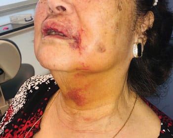
Common Symptoms: Most published case reports describe similar sequelae following an NaOCl accident — sudden pain, profuse bleeding and immediate swelling (Figure 1). This triad of signs and symptoms can be considered pathognomonic for an NaOCl extrusion event.17
Risk Factors: The anatomical position of the root apex relative to the alveolar bone is critical, especially when the apex of the root is only surrounded by a layer of thin bone or solely by soft tissue. In these cases, the extrusion of even a small volume of NaOCl may lead to severe symptoms and the potential to spread through the soft tissue and fascial planes. Other teeth at a higher risk for extrusion injuries include those with cortical bone fenestration, an open sinus tract, or open communication with the maxillary sinus. Bone density appears to play an important role, since extrusion injuries are more common in the maxilla compared to the mandible, and more common in females than males.16
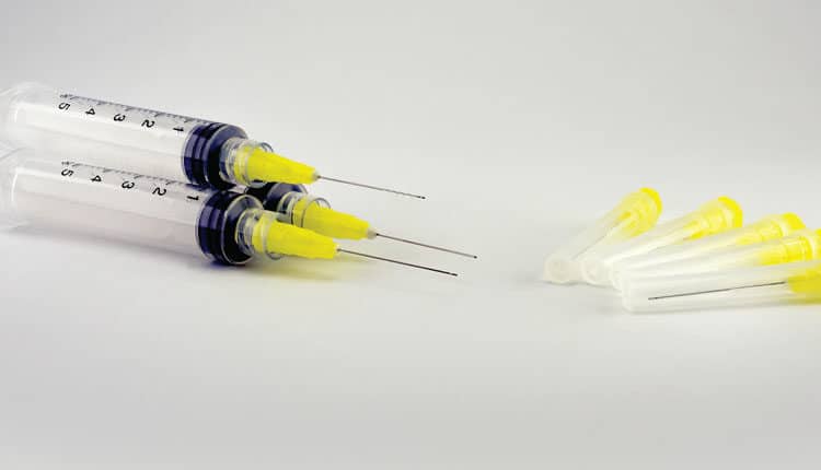
Over-enlargement of the apical foramen or an iatrogenic perforation are additional avenues for NaOCl extrusion. Other factors include immature teeth with wide apical foramina or teeth presenting with perforating internal or external resorption. Inappropriate irrigation techniques (e.g., wedging the irrigation needle in the root canal) can also result in excessive forces which can push NaOCl into the periapical tissues. Using a needle with an open-ended tip, as opposed to a side-vented needle, has also been found to be a contributing factor in NaOCl extrusion accidents.17
Prevention and Management: Although there are no standardized guidelines, there have been several published recommendations for the management of an NaOCl accident.18,19 Fortunately, all or most of the signs and symptoms resolve within a few weeks in the majority of cases.
Prevention
- Utilize preoperative radiographs and cone beam computed tomography to assess the root canal anatomy and root proximity to vital structures, such as the inferior alveolar nerve, mental foramen or sinus.
- Use side-vented irrigation needles.
- Irrigate at least 2 mm short of the working length.
- Do not wedge the needle tip in the canal.
- Use constant and gentle in-and-out movements of the needle during irrigation.
Managing an NaOCl Extrusion Accident
- Immediately irrigate the canal with normal saline to dilute the NaOCl.
- Allow bleeding to occur through the canals.
- Advise patients to use cold compression for the first 24 hours (15 minutes at a time) to minimize swelling.
- Recommend warm compression after 24 hours (15 minutes at a time) for 24 hours.
- Prescribe analgesics for seven days (if not medically contraindicated).
- Prescribe antibiotic coverage to prevent secondary infection, as well as steroid therapy.
- Reassure the patient and provide the individual with both verbal and written home care instructions.
- Monitor the patient daily.
- If needed, refer the patient to an oral surgeon or to an emergency department.
- Document the event, its management, and patient instructions thoroughly in the patient’s record.
CALCIUM HYDROXIDE EXTRUSION
Prevalence: The first case report of an adverse reaction to a Ca(OH)2 extrusion was published in 2000. Since then, several more case reports have been published.20 Compared to the incidence of NaOCl injuries, Ca(OH)2 extrusion injuries occur even less frequently. However, like NaOCl extrusion accidents, the actual number of Ca(OH)2 extrusion accidents is most likely underestimated due to the large number of accidents that are neither reported nor reach publication.
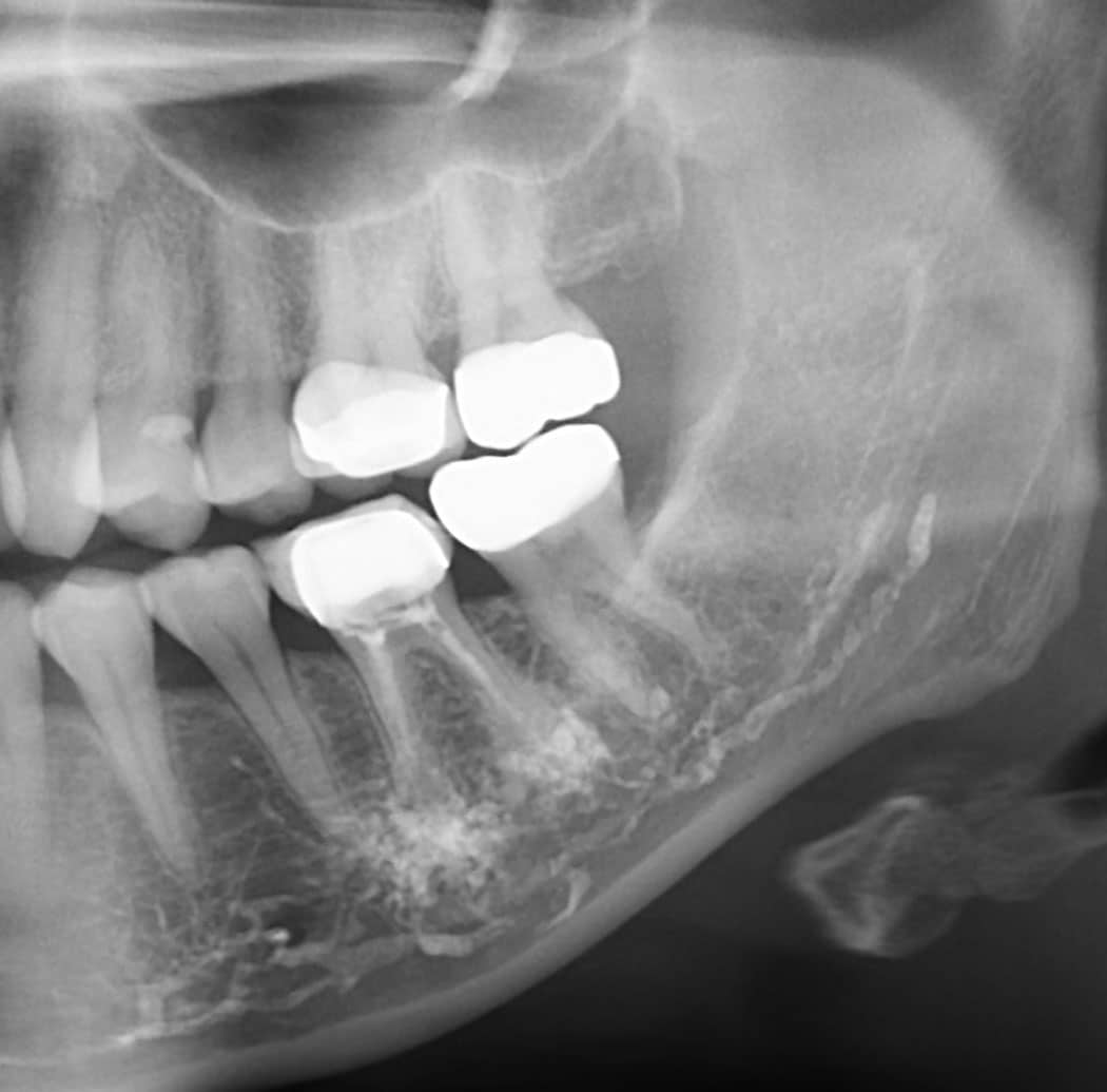
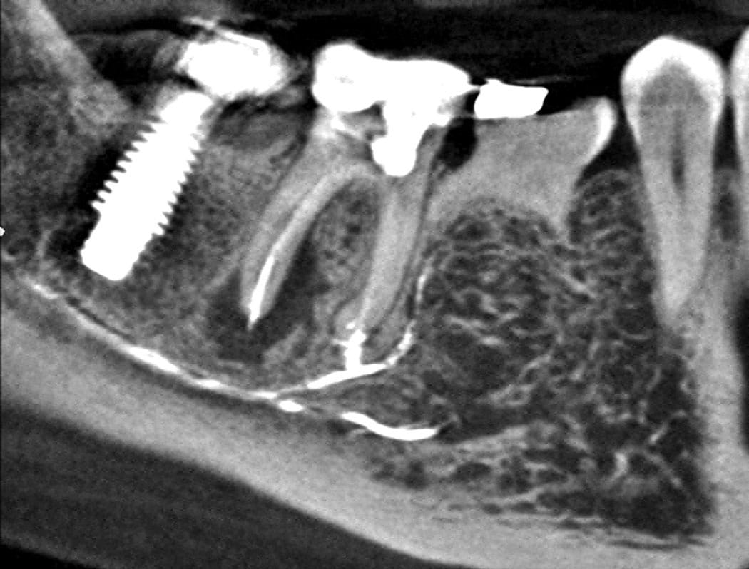
Common Symptoms: Extrusion of Ca(OH)2 can cause chemical injury, compressive injury or both when the material is expressed into the confines of a space that is only meant to be occupied by the neurovascular bundle (Figure 2 and Figure 3).21 The host reaction to Ca(OH)2 extrusion into the soft tissue or blood vessels can range from a minor inflammatory reaction to serious sequelae, including tissue necrosis and paresthesia. This depends on the amount of material that is extruded into the periapical region and volume of material in contact with vascular and neural tissues.21
Risk Factors: Several case reports have identified potential causes for Ca(OH)2 extrusion. The overall risk was found to be higher when low-viscosity Ca(OH)2 pastes were used in combination with injection under pressure and the use of non-side-vented irrigation needles. Another potential cause was the needle binding in the canal during Ca(OH)2 delivery, which resulted in excessive pressure. Other factors included over-enlargement of the apical constriction during instrumentation, teeth with an immature apex, or a tooth with a resorptive defect.
The amount of Ca(OH)2 that is extruded through the apical foramen during syringe delivery depends on the viscosity of Ca(OH)2 used, type of needle (open-ended versus side-vented), and needle diameter. The depth of the needle placement also plays an important role, as does the final dimensions of the apical terminus and/or prepared canal taper. Lastly, the operation of the syringe, as well as the injection pressure, can be contributing factors in Ca(OH)2 extrusion accidents.21
Prevention and Management: Adopting the following approach will help clinicians avoid or manage iatrogenic extrusions during root canal treatment.
Prevention
The following are some reasonable and responsible recommendations to follow when using Ca(OH)2 as an interim medicament, based on previous published guidelines:21
- Assess radiographs and cone beam images to identify if teeth are in close proximity to the inferior alveolar nerve or sinuses.
- Take special care to prevent over-enlargement of the apices of mandibular premolar and molar teeth, which can contribute to the extrusion of materials.
- Consider the use of a Lentulo spiral as a safer alternative to syringe needle delivery. A recent in vitro study showed that a Lentulo spiral filler at 500 rpm, 3 mm short of the apex, minimized extrusion of Ca(OH)2from root canals.22
- Paper point application is safe alternative to injection delivery.
- Follow the manufacturer’s guidelines when using a commercial prefilled Ca(OH)2syringe.
- Make sure the needle does not bind in the canal when injecting.
- Use a slow injection rate and constant outward movement from the canal as the material is injected.
- Take appropriate postoperative periapical radiographs to check for any extrusion of dressing or filling materials into the inferior alveolar canal, around the mental foramen, or near other vital structures.
Managing a Ca(OH)2 Extrusion Accident
- Document events thoroughly and refer the patient to an oral surgeon or endodontist for follow-up.
- If the patient reports any postoperative paresthesia, dysesthesia or numbness within the first 24 to 72 hours, time is critical in this true neurologic emergency, and the patient should be referred to an oral surgeon for possible debridement.
Immediate Assessment
The literature recommends an immediate assessment of symptoms and thorough discussion with the patient on the timing and prognosis for a surgical intervention to remove the extruded material. When warranted after a specialist consultation, the extruded material should be removed surgically if neurovascular damage is suspected.
Studies have shown that in cases in which a material was injected into the inferior alveolar nerve space that resulted in a neurosensory injury, expedient management of the extrusion site through decompression and debridement significantly improved the prognosis.23–25 In one study of 61 patients who experienced an endodontic sealer extrusion into the inferior alveolar nerve canal, 11 patients underwent surgical exploration. The five patients who underwent surgical decompression and debridement within the first 48 hours all recovered completely. The remaining six patients underwent surgical exploration between 10 days and three months after receiving endodontic therapy. Among these patients, four experienced partial recovery and two experienced no recovery.26
CONCLUSION
In conclusion, although rare, dental practitioners should be aware of the possible risks that commonly used irrigants and intracanal medicaments can cause if accidentally extruded beyond the apex. Prompt realization and appropriate actions are necessary to avoid any serious complications and long-term damage.
References
- Tronstad L. Recent development in endodontic research. Scand J Dent Res. 1992;100:52–59.
- Siqueira JF. Aetiology of root canal treatment failure: why well-treated teeth can fail. Int Endod J. 2001;34:1–10.
- Siqueira JF, Araujo MCP, Garcia PF, Fraga RC, Dantas CJ. Histological evaluation of the effectiveness of five instrumentation techniques for cleaning the apical third of root canals. J Endod. 1997;23:499–502.
- Wu MK, Dummer PM, Wesselink PR. Consequences of and strategies to deal with residual post-treatment root canal infection. Int Endod J. 2006;39:343–356.
- Peters OA, Schonenberger K, Laib A. Effects of four Ni-Ti preparation techniques on root canal geometry assessed by micro computed tomography. Int Endod J. 2001;34:221–230.
- Zehnder M. Root canal irrigants. J Endod. 2006;32:389–398.
- Turkun M, Cengiz T. The effects of sodium hypochlorite and calcium hydroxide on tissue dissolution and root canal cleanliness. Int Endod J. 1997;30:335–342.
- Sjögren U, Figdor D, Spångberg L, Sundqvist G. The antimicrobial effect of calcium hydroxide as a short‐term intracanal dressing. Int Endod J. 1991;24:119–125.
- Hasselgren G, Olsson B, Cvek M. Effects of calcium hydroxide and sodium hypochlorite on the dissolution of necrotic porcine muscle tissue. J Endod. 1988;14:125–127.
- Safavi KE, Nichols FC. Effect of calcium hydroxide on bacterial lipopolysaccharide. J Endod. 1993;19:76–78.
- Peters LB, van Winkelhoff AJ, Buijs JF, Wesselink PR. Effects of instrumentation, irrigation and dressing with calcium hydroxide on infection in pulpless teeth with periapical bone lesions. Int Endod J. 2002;35:13–21.
- Olsen JJ, Thorn JJ, Korsgaard N, Pinholt EM. Nerve lesions following apical extrusion of non-setting calcium hydroxide: a systematic case review and report of two cases. J Craniomaxillofac Surg. 2014;42:757–762.
- Pashley EL, Birdsong NL, Bowman K, Pashley DH. Cytotoxic effects of NaOCl on vital tissue. J Endod. 1985;11:525–528.
- Swanljung O, Vehkalahti MM. Root canal irrigants and medicaments in endodontic malpractice cases: a nationwide longitudinal observation. J Endod. 2018;44:559–564.
- Boutsioukis C, Psimma Z, van der Sluis LW. Factors affecting irrigant extrusion during root canal irrigation: a systematic review. Int Endod J. 2013;46:599–618.
- Kleier DJ, Averbach RE, Mehdipour O. The sodium hypochlorite accident: experience of diplomates of the American Board of Endodontics. J Endod. 2008;34:1346–1350.
- Guivarc’h M, Ordioni U, Ahmed HMA, Cohen S, Catherine J-H, Bukiet F. Sodium hypochlorite accident: a systematic review. J Endod. 2017;43:16–24.
- Farook SA, Shah V, Lenouvel D, Sheikh O, Sadiq Z, Cascarini L. Guidelines for management of sodium hypochlorite extrusion injuries. Br Dent J. 2014;217:679–684.
- Spencer H, Ike VDV, Brennan PA. Review: the use of sodium hypochlorite in endodontics — potential complications and their management. Br Dent J. 2007;202:555–559.
- Al-Sheeb F, Al Mannai G, Tharupeedikayil S. Nicolau syndrome after endodontic treatment: a case report. J Endod. 2022;48:269–272.
- Gluskin AH, Lai G, Peters CI, Peters OA. The double-edged sword of calcium hydroxide in endodontics: precautions and preventive strategies for extrusion injuries into neurovascular anatomy. J Am Dent Assoc. 2020;151:317–326.
- Lai GS, Davis S, Gluskin AH, Peters CI, Peters OA. Comparison of calcium hydroxide extrusion with syringe versus spiral filler delivery: A pilot study. Aust Endod J. 2021;47:408–414.
- Grotz KA, Al-Nawas B, de Aguiar EG, Schulz A, Wagner W. Treatment of injuries to the inferior alveolar nerve after endodontic procedures. Clin Oral Investig. 1998;2:73–76.
- Ahlgren FK, Johannessen AC, Hellem S. Displaced calcium hydroxide paste causing inferior alveolar nerve paraesthesia: report of a case. Oral Surg Oral Med Oral Pathol Oral Radiol Endod. 2003;96:734–737.
- Giuliani M, Lajolo C, Deli G, Silveri C. Inferior alveolar nerve paresthesia caused by endodontic pathosis: a case report and review of the literature. Oral Surg Oral Med Oral Pathol Oral Radiol Endod. 2001;92:670–674.
- Pogrel MA. Damage to the inferior alveolar nerve as the result of root canal therapy. J Am Dent Assoc. 2007;138:65–69.
From Decisions in Dentistry. February 2023;9(2)xx.
