
Etiology and Management of White Spot Lesions
Determining the cause of these lesions is essential to successful treatment and esthetic improvement.
Determining the cause of these lesions is essential to successful treatment and esthetic improvement.
The presence of clinically detectable, localized areas of enamel demineralization, observed as white spot lesions of different opacity, is a sign that the caries process has begun. Dental caries results in the dissolution of apatite crystals and the loss of calcium, phosphate and other ions, which eventually leads to demineralization of the tooth substrate.1 The subsurface porosity caused by demineralization gives the lesion a milky appearance that can be found on the smooth surfaces of teeth.2
White spot lesions are not only the result of demineralization, however, as fluorosis, hypomineralization/hypomaturation and hypoplasia can also cause lesions. Dental professionals are charged with performing a differential diagnosis to determine the etiology of white spot lesions, as well as providing appropriate treatment and esthetic management that will meet patients’ expectations.
While fluoride remains an important factor in the prevention and management of dental caries, widespread exposure from different sources has increased the risk of fluorosis in communities, regardless of whether or not the community uses a fluoridated water supply.3 Unlike early caries lesions, dental fluorosis is a developmental disturbance caused by exposure to high concentrations of fluoride during tooth development, which leads to enamel with lower mineral content and increased porosity.4 Fluorosis may present initially as white spot lesions that progress to brown spots, due to the inherent porosity that makes them susceptible to stain.
Another form of developmental enamel anomaly that leads to discoloration is associated with genetic defects and environmental insults, such as metabolic conditions and exposure to drugs, chemicals, radiation and trauma.5 These defects are associated with hypoplasia, hypomineralization or hypomaturation. Enamel hypoplasia occurs if the matrix formation is affected and results in pits or grooves, or thin and missing enamel. Hypomineralization is due to maturation disturbance, which results in reduced mineralization and commonly presents as soft enamel. Hypomaturation is caused by a reduction in the deposition of minerals at the end stage of mineralization, and is characterized by altered translucency affecting the entire tooth. Or, if noted in a localized area, it presents as an opacity.6
COURTESY OF MARCOS VARGAS, BDB, DDS, MS
COURTESY OF MARCOS VARGAS, BDB, DDS, MS
DIFFERENTIAL DIAGNOSIS
Considering the various etiologies of white spot lesions, it is imperative to establish a proper diagnosis. This is based on a thorough review of dental and medical history, and clinical examination evaluating the location, symmetry, outline form, depth and opacity of the lesion. The information gathered must include a history of neonatal or early childhood illness, use of drugs and medications, and past infections or trauma related to primary teeth.7
Fluoride exposure is another important factor in the diagnosis. Patients should be asked whether they reside in a community with water fluoridation. They should also be asked about any history of fluoride supplementation, the amount of toothpaste used, and history of swallowing toothpaste in childhood.7 While the criteria for dental fluorosis is clear, there are circumstances in which a final diagnosis may be challenging. Additionally, individual caries risk assessments should be performed so the most appropriate intervention can be implemented before permanent damage is done to tooth structure.
The lesion’s descriptive clinical presentation is another important guide in the diagnostic process. Several classifications and indices are used: the Al-Alousi Index,8 Developmental Defects of Enamel Index (DDE),9 and Modified DDE Index.6 According to the Modified DDE Index, developmental defects are differentiated based on three broad visual clinical categories: demarcated opacities, diffuse opacities and hypoplasia.
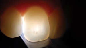
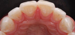
The extent of the defect is also considered. When selecting treatment options, for example, lesion depth is a critical component that can be estimated with the aid of a transilluminator. The transilluminator is positioned on the lingual surface to examine the lesion depth, as a darker color indicates deeper staining (Figure 1).
Demineralization and white spots associated with early noncavitated lesions can be observed on smooth surfaces of teeth along the gingiva and interproximal areas. Based on the International Caries Detection and Assessment System, if no lesion is apparent on a smooth surface when the tooth is wet, but a noncavitated lesion appears once the tooth is adequately dried, the lesion is classified as a Code 1. If a noncavitated lesion is apparent both when the tooth is wet or dry, it is classified as a Code 2.10 The outline is usually well-defined, and follows the contour of the gingival tissue. Upon identification of demineralization, it is important to assess the lesion activity to properly plan treatment. Predictors of an active lesion include: located in a plaque stagnation area, white in color, dull appearance, and presence of rough surface or surface breakdown. Predictors of an inactive or arrested lesion include: located in a nonplaque stagnation area (above the gingival margin), brown in color, shiny appearance, and intact, smooth and hard surface.11
Dental fluorosis commonly occurs bilaterally and presents with a thin, diffuse and lattice-type opacity corresponding to perikymata running across the tooth surface to an entirely chalky white overall tooth surface.12 The lesion can be localized to a few teeth or can include the full dentition. The lesion’s depth can vary from superficial to deep into the enamel, based on the severity of the fluorosis.
Hypomineralization and hypomaturation lesions present with well-demarcated margins — usually affecting one or several teeth — with symmetrical distribution.9 Hypoplasia is characterized by diffuse opacities of white and yellow discoloration, often accompanied with pitting and loss of enamel structure. Slightly different clinical representations of white spot lesions may occur simultaneously.
key takeaways
- White spot lesions of different etiologies can cause functional and esthetic concerns, and are not only the result of demineralization. Fluorosis, hypomineralization/ hypomaturation and hypoplasia can also cause these lesions.
- Considering the various etiologies of white spot lesions, it is imperative to establish a proper diagnosis. Upon identification of demineralization, clinicians must assess lesion activity in order to plan proper treatment.
- Minimal intervention is an ideal approach in managing white spot lesions, and should start with remineralization therapies.
- If a white spot lesion is an esthetic concern, tooth whitening should be considered in an attempt to blend the lesion in with the natural dentition.
- Restorative treatment is indicated when conservative approaches — such as remineralization, tooth whitening, microabrasion and resin infiltration — are unsuccessful in removing or masking the white spot lesion.
MANAGING LESIONS
Minimal intervention is an ideal approach in managing white spot lesions, and should start with remineralization therapies to arrest the disease process and restore enamel strength and function. The patient’s expectations are vital to the decision-making process, as esthetic concerns should be addressed concurrently. No treatment may also be an option.
Ideally, demineralization should be prevented by reducing the frequency of fermentable carbohydrate consumption, decreasing the number of cariogenic microbes present, and increasing salivary flow. The best evidence for remineralization suggests topical use of fluoride, based on the patient’s caries risk assessment. In a high-risk patient, a prescription 5000 ppm fluoride dentifrice may be recommended, in addition to the in-office application of 2.26% fluoride varnish.13 Silver diamine fluoride has recently become available in the United States, and application of this liquid agent can arrest active dentinal and enamel caries, preventing further progression of the disease.14,15 But because silver diamine fluoride stains the lesions that it arrests, its use on permanent anterior teeth may be contraindicated. Calcium phosphate technologies, including amorphous calcium phosphate (ACP), casein phosphopeptide-ACP, calcium sodium phosphosilicate, and tricalcium phosphate, are also available, although additional research is needed to support their use.11
ESTHETIC CONCERNS
If a white spot lesion is an esthetic concern, tooth whitening should be considered in an attempt to blend the lesion in with the natural dentition. Tooth whitening involves diffusion of the whitening material into the enamel and dentin to interact with stain molecules. It also creates micromorphologic alterations on the tooth surface that affect its optical properties.16 When properly monitored by a dental professional, tooth whitening is safe and effective in improving the appearance and color of teeth.17
Tooth whitening with hydrogen peroxide or carbamide peroxide can be broadly classified into three categories: in-office whitening; professionally supervised take-home treatment; and over-the counter whitening. In-office whitening is performed using highly concentrated materials of up to 40% hydrogen peroxide, and is frequently combined with light activating devices. Prescription at-home whitening systems use custom-fabricated trays and carbamide peroxide gels (in concentrations ranging from 7% to 22%). Over-the-counter whitening products are available in strips, paint-on gels or brush-on adhesive liquids. Current research on the efficacy of in-office whitening versus at-home whitening shows that both techniques provide satisfactory outcomes without significant adverse effects in terms of tooth and gingival sensitivity.18

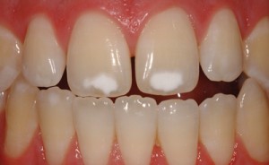
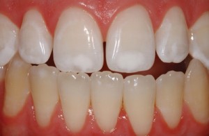
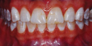
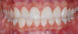
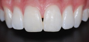
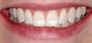
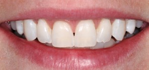
In many instances, whitening will lighten the tooth color so that the white spot lesion naturally blends in (Figure 2A and Figure 2B). Upon completion of the whitening process, patients can decide if further treatment, such as minimally invasive restorative procedures, is needed to meet their esthetic expectations.
Enamel microabrasion is indicated for intrinsic discoloration or texture alteration due to enamel hypoplasia, amelogenesis imperfecta or fluorosis.19 It involves a combination of erosion and abrasion of the superficial enamel by a low concentration of hydrochloric acid, in conjunction with mechanical rubbing of abrasive silicon carbide particles in a water-soluble mixture.20 The success of microabrasion is related to proper case selection and precise technique. Microabrasion is used for superficial defects, but is contraindicated in lesions with dentin involvement and deeper opaque stains associated with severe hypoplasia, which may require a restorative approach.21
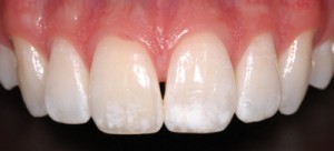
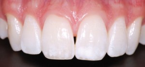
When overall tooth color change is desired, microabrasion can be preceded or followed by tooth whitening to improve the overall esthetic outcome (Figure 3A and Figure 3B). Proper isolation with a rubber dam is necessary during microabrasion procedures to ensure patient safety. Furthermore, periodic evaluation of the enamel thickness labio-lingually with a mouth mirror is helpful in assessing the amount of enamel removed during the procedure (Figure 4A through Figure 4E). Upon completion of microabrasion, fluoridated prophylaxis paste should be applied to aid remineralization of the treated surface.
COURTESY OF MARCOS VARGAS, BDB, DDS, MS
COURTESY OF MARCOS VARGAS, BDB, DDS, MS
Resin infiltration with a low-viscosity hydrophilic resin material in the porous lesion has been proposed as a treatment for white spot lesions.22 The idea is that further demineralization can be inhibited by the infiltration of the matrix into the lesion. An additional benefit is that the refractive index of the lesion is altered to make it comparable to that of the natural tooth, thereby enhancing esthetics.
Resin infiltration consists of 15% hydrochloric acid with a proprietary resin infiltrate. After isolation, the white spot lesion is etched with 15% hydrochloric acid for two minutes, followed by rinsing and drying with ethanol. The last step involves the application of the resin infiltrate, which is then light cured. Because this technique was only recently introduced, long-term results on the efficacy and stability of resin infiltration in the treatment of white spot lesions are not available. A short-term clinical study found that the esthetic outcome with resin infiltration was satisfactory for the treatment of post-orthodontic white spots.23 A combination of microabrasion followed by resin infiltration can also be used to improve the esthetic outcome of white spot lesions (Figure 5A and Figure 5B).
Restorative treatment with direct resin composites is indicated when conservative approaches — such as remineralization, tooth whitening, microabrasion and resin infiltration — are unsuccessful in removing or masking the white spot lesion. This is often seen in deeper lesions associated with enamel hypoplasia; in these cases, the lesion typically has to be removed and the lost tooth structure replaced with a composite restoration.7
CONCLUSION
White spot lesions of different etiologies can cause functional and esthetic concerns that must be managed properly to meet patients’ expectations. Careful review of medical and dental histories and a detailed clinical examination will aid in the differential diagnosis of superficial white spot lesions. Treatment planning and management should emphasize proper sequencing, starting with the most conservative treatment option. The patient should also be part of the decision-making process from start to finish.
ACKNOWLEDGEMENT
The authors would like to thank Marcos Vargas, BDB, DDS, MS; Marcela Hernandez, DDS, MS; and Justine Kolker, DDS, MS, PhD, for their help with this manuscript.
References
- Cochrane NJ, Cai F, Huq NL, Burrow MF, Reynolds EC. New approaches to enhanced remineralization of tooth enamel. J Dent Res. 2010;89:1187–1197.
- Mizrahi E. Enamel demineralization following orthodontic treatment. Am J Orthod. 1982;82:62–67.
- Iida H, Kumar JV. The association between enamel fluorosis and dental caries in US schoolchildren. J Am Dent Assoc. 2009;140:855–862.
- Abanto Alvarez J, Rezende KM, et al. Dental fluorosis: exposure, prevention and management. Med Oral Patol Oral Cir Bucal. 2009;14:103–107.
- Seow WK. Developmental defects of enamel and dentin: challenges for basic science research and clinical management. Aust Dent J. 2014;59:143–154.
- Clarkson J. Review of terminology, classification, and indices of developmental defects of enamel. Adv Dent Res. 1989;3:104–109.
- Wray A, Welbury R. UK National Clinical Guidelines in Paediatric Dentistry: Treatment of intrinsic discoloration in permanent anterior teeth in children and adolescents. Int J Paediatr Dent. 2001;11:309–315.
- Al-Alousi W, Jackson D, Compton G, Jenkins OC. Enamel mottling in a fluoride and in a non-fluoride community. Br Dent J. 1975;138:56–60.
- An epidemiological index of developmental defects of dental enamel (DDE Index). Commission on Oral Health, Research and Epidemiology. Int Dent J. 1982;32:159–167.
- ICDAS II — International Caries Detection and Assessment System (ICDAS II) Criteria Manual. Available at: icdas.org/uploads/ICDAS%20Criteria %20Manual%20Revised%202009_2.pdf. Accessed December 10, 2015.
- Kwon SR, Kolker J. Implement a minimally invasive approach. Dimensions of Dental Hygiene. 2014;12(8):22–25.
- Fejerskov O, Kidd E. Dental Caries — The Disease and its Clinical Management. 2nd ed. Hoboken, New Jersey: Wiley-Blackwell; 2008.
- American Dental Association Guide: topical fluoride for caries prevention: executive summary of the updated clinical recommendations and supporting systematic review. J Am Dent Assoc. 2013;144:1279–1291.
- Peng JJ, Botelho MG, Matinlinna JP. Silver compounds used in dentistry for caries management: a review. J Dent. 2012;40:531–541.
- Beltrán-Aguilar ED. Silver diamine fluoride (SDF) may be better than fluoride varnish and no treatment in arresting and preventing cavitated carious lesions. J Evid Based Dent Pract. 2010;10:122–124.
- Kwon SR, Wertz PW. Review of the mechanism of tooth whitening. J Esthet Restor Dent. 2015;27(5):240–257.
- American Dental Association Council on Scientific Affairs. Tooth Whitening/Bleaching: Treatment Considerations for Dentists and Their Patients. Chicago: American Dental Association; 2009.
- Auschill TM, Hellwig E, Schmidale S, Sculean A, Arweiler NB. Efficacy, side-effects and patients’ acceptance of different bleaching techniques (OTC, in-office, at-home). Oper Dent. 2005;30:156–163.
- Sunfeld RH, Croll TP, Briso AL, de Alexandre RS, Sundfeld Neto D. Considerations about enamel microabrasion after 18 years. Am J Dent. 2007;20:67–72.
- Croll TP, Helpin ML. Enamel microabrasion: a new approach. J Esthet Dent. 2000;12:64–71.
- Reston EG, Corba DV, Ruschel K, Tovo MF, Barbosa AN. Conservative approach for esthetic treatment of enamel hypoplasia. Oper Dent. 2011;36:340–343.
- Paris S, Meyer-Lueckel H. Inhibition of caries progression by resin infiltration in situ. Caries Res. 2010;44:47–54.
- Knösel M, Eckstein A, Helms HJ. Durability of esthetic improvement following Icon resin infiltration of multibracket-induced white spot lesions compared with no therapy over 6 months: a single-center, split-mouth, randomized clinical trial. Am J Orthod Dentofacial Orthop. 2013;144:86–96.
