
Intraoral Scanning in the Creation and Application of Duplicate Dentures
This case report illustrates the clinical use of intraoral scanners in the fabrication of duplicate dentures.
Intraoral scanners have proven to be clinically useful in digital guided implant surgery. Aside from digital guided surgery, intraoral scanners are also used for the fabrication of duplicate dentures via denture scanning. In various cases of actual clinical practice, patients may need to replicate previously used complete dentures.
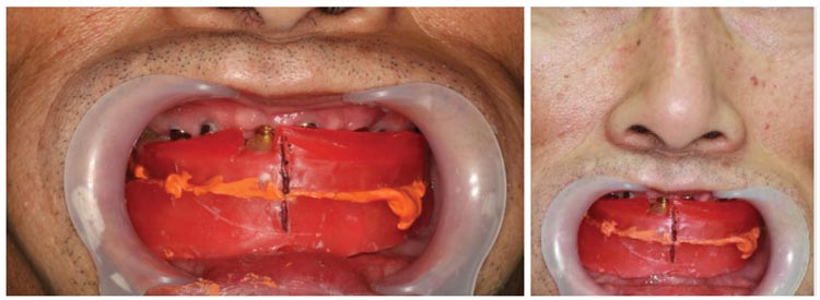
When complete dentures are used as temporary prosthetics after implant placement, wax rims are normally used to obtain a final impression and then to convey information about the patient’s jaw relationship and vertical dimension to the laboratory (Figure 1). A wax rim is connected to three or four implants to ensure safety, and this process causes patient discomfort. Also, after impression taking, another visit is required to acquire the vertical dimension and jaw relationship.
Advantages of Intraoral Scanning
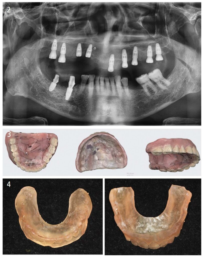
In this case, the inner and outer surfaces of the temporary complete denture used after implant surgery can be scanned using an intraoral scanner, which can then be printed using a 3D printer to easily create duplicate dentures at the clinic or laboratory (Figure 2 and Figure 3).
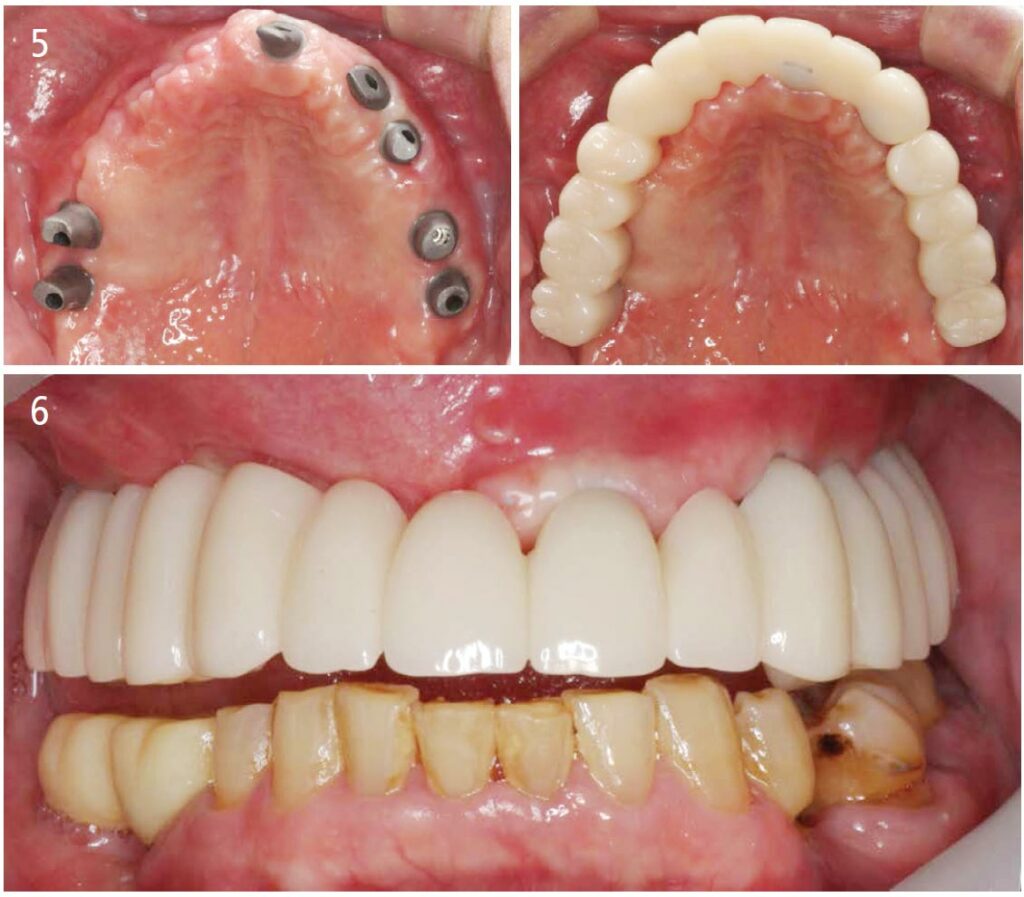
The maxillary and mandibular models can be mounted in the laboratory using the duplicate denture, reducing the number of visits and chairtime for the patient (Figure 4 through Figure 6).
In addition to the prosthetic restoration of implants, scanning dentures with intraoral scanners to duplicate existing dentures can reduce total treatment time and the number of visits needed.
Conclusion
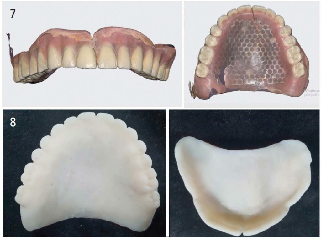
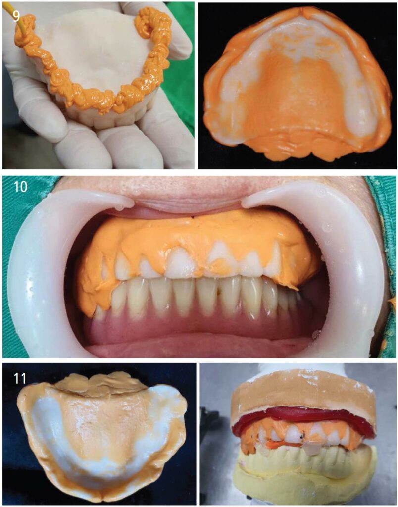
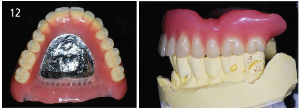
As seen in this case, creating duplicate dentures using intraoral scanners (Figure 7 and Figure 8) eliminates the need for a preliminary impression to make an individual tray and the wax rim stage, reducing the number of patient visits and chairtime (Figure 9 through Figure 11). In addition, using a wireless intraoral scanner for denture scans removes the distance limit from a PC and offers more freedom of operation for increased ease and reduced burden on the wrist. And with the same scanning speed as a wired intraoral scanner, it can also reduce overall work time in completing the final denture (Figure 12).
Recently, the adoption rate of intraoral scanners has been increasing rapidly, but the reality is that many clinicians don’t have a set plan for how to use them. Aside from prosthetic and restorative treatments, I hope my use of intraoral scanners may be of some help to readers.
Medit
i700wireless.com
From Decisions in Dentistry. June 2022;8(6)22,24.
