

Applications and Limitations of Periodontal Endoscopy
An examination of the use of this technique in nonsurgical periodontal therapy.
An examination of the use of this technique in nonsurgical periodontal therapy
In 2015, the American Dental Association (ADA) Council on Scientific Affairs released a systematic review and meta-analysis of nonsurgical treatment for patients with chronic periodontitis. The report concluded that conventional scaling and root planing, with or without adjuncts, should be considered the initial treatment modality for patients presenting with this chronic disease.1

ERAXION/ISTOCK/ THINKSTOCK
The primary objective of scaling and root planing is to restore periodontal health by removing pathogenic materials and products (e.g., biofilm, calculus and endotoxins) that induce inflammation from periodontally involved root surfaces. Calculus has been shown to contain bacterial products that contribute to the inflammatory response and can perpetuate periodontal infection.2–4 Numerous studies, however, indicate clinical difficulty in determining the thoroughness of subgingival instrumentation, as well as clinical limitations in instrumenting subgingival root surfaces in a closed pocket, particularly in pocket depths greater than 5 mm.5–8
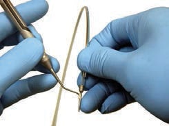
The periodontal endoscope facilitates visualization of the subgingival environment via a 1-mm-diameter, flexible fiber-optic probe with imaging capabilities (Figure 1). This fiber-optic system enlarges the image while also delivering light to the working field.
ERAXION/ISTOCK/ THINKSTOCK
In use, the endoscopic fiber is covered with a single-use, sterile endoscopic sheath.9 The disposable sheath provides a barrier to pathogens, preventing cross contamination. The sheath protects the fiber and bonded sapphire lens (housed inside a metal cannula), and is designed to deliver water irrigation, keep the endoscope lens clear, and eliminate the need for disinfection of the fiber between patients.
The fiber/sheath/explorer complex (Figure 2) is placed into the sulcus by the clinician for subgingival viewing.10 The real-time video image is displayed on a chairside monitor and may be magnified between 24 and 48 times, depending on the proximity of the fiber-optic lens to the area being viewed. Table 1 summarizes the advantages and disadvantages of this technique.
An early study by Stambaugh et al evaluated the periodontal endoscope and the ability of the clinician to develop skills in using fiber-optics to accurately visualize the contents of the subgingival sulcus.11 Results from other studies demonstrated that adjunctive use of the periodontal endoscope for nonsurgical periodontal therapy resulted in improved visibility of deposits for calculus removal.4,12
Wilson et al found that root surfaces treated endoscopically with a single course of closed subgingival scaling and root planing demonstrated no histologic signs of chronic inflammation six months after therapy.13
 Clinical Applications of the periodontal Endoscope
Though not a comprehensive list, uses for periodontal endoscopy include:
- Patients with probing depths greater than 4 mm who are beginning initial periodontal therapy
- Individuals with sites that have not responded to traditional nonsurgical debridement
- Periodontal maintenance patients with chronic inflammation or increasing probing depths
- Residual probing depths in periodontal maintenance patients who refuse surgical therapy, and/or where surgery is contraindicated for medical or esthetic reasons
- Patients with suspected subgingival pathology, such as root fractures, endodontic root perforations or resorption
- Other subgingival pathologies identified with this technology include caries, open restoration margins and cervical enamel projection
Endoscopic root debridement is achieved using hand instruments or ultrasonic inserts or tips (UITs); this allows for microvisual instrumentation and optimizes subgingival healing. The following periodontal endoscopic cases are consistent with the 2015 ADA Council on Scientific Affairs recommendations that conventional scaling and root planing, with or without adjuncts, be considered the initial treatment for patients with chronic periodontitis.1
Elemental iodine or its derivatives (povidone-iodine) may be the most broad spectrum and potent antiseptics available.17 Elemental iodine’s broad spectrum antimicrobial activity, low potential for developing resistance and adverse reactions, wide availability, ease of use, and low cost make it a valuable adjunct to periodontal therapy.18
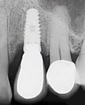
In a 2009 study, Wilson found that excess dental cement was associated with signs of peri-implant disease.19 One possible explanation is that cement retains microbes, similar to those responsible for inflammatory periodontal disease, and the rough surface of the cement inhibits the removal of the microorganisms, which can lead to peri-implant disease. The study noted that after removal of excess cement, clinical and endoscopic signs of peri-implant disease were absent at 30 days posttreatment.19 A later study found small pieces of cement in the soft tissues, however, which may warrant flap surgery in some cases.20
Even the most diligent clinicians may sometimes miss residual subgingival cement in implant cases. For this reason, maintenance is extremely important for patients who have cement-retained implant restorations. Signs of peri-implant inflammation often serve as a warning that there is excess cement below the gumline, and alert clinicians before there is radiographic bone loss. Other symptoms can include localized inflammation, progressively deeper probing depths, or bleeding on probing. If these signs are caught early, it may be possible to remove excess cement before irreversible damage occurs.21
The following cases show how endoscopic debridement of an implant with retained cement may resolve once the cement is microscopically removed.
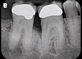
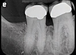
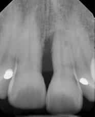
Case 1: Endoscopic debridement #30 distal, no adjuncts used. Site maintained on three-month supportive periodontal therapy (figures 3A and 3B)
Treatment by Janell Hall, RDH, in the periodontal office of Nicholas Missichia, DDS, MS, Bend, Oregon
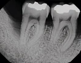
Case 2: Endoscopic debridement #31 distal, with adjunctive microencapsulated minocycline (figures 4A and 4B)
Treatment by Deborah Murchison, RDH, in the periodontal office of John Y. Kwan, DDS, Oakland, California
Micro-encapsulated minocycline is a locally delivered drug that the clinician places into diseased sites in the periodontium. The drug releases the antimicrobial at a steady pharmacological level for an extended period.14

Case 3: Endoscopic debridement #3 distal, with the nonconcurrent use of adjunctive antibiotic amoxicillin and local antibiotic administration of micro-encapsulated minocycline (figures 5A and 5B)
Treatment by Sarah Bacich, RDH, in the periodontal office of David Trylovich, DDS, MS, Las Vegas, Nevada


Case 4: Endoscopic debridement #9 mesial, with adjunctive systemic antibiotic amoxicillin and metronidazole (figures 6A and 6B)
Treatment by Amy Curran, RDH, in the periodontal office of Ronald Watkins, DDS, MS, Phoenix, Arizona
Periodontal lesions often harbor a mixture of pathogenic bacteria. As a result, drug combination therapies have gained increasing importance. In fact, they may even be required for eradication and prevention of periodontal infections by known periodontal pathogens that invade subepithelial periodontal tissue or colonize extra-dental domains — from which they may translocate to periodontal sites.15
Antibiotic combination therapy with two antibiotics can be used to take advantage of different mechanisms of action and to expand the spectrum of antimicrobial activity. Amoxicillin-metronidazole (250 mg amoxicillin; 375 mg metronidazole, three times daily for eight days) is the most common antibiotic combination in periodontics.16

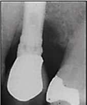
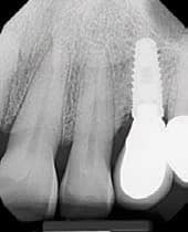
Case 5: Endoscopic debridement implant #12 presenting with periimplant disease, with adjunctive systemic antibiotic amoxicillin and metronidazole (figures 7A and 7B)
Treatment by John Y. Kwan, DDS

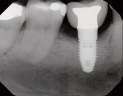
Case 6: Endoscopic debridement implant #5 presenting with periimplant disease, with adjunctive systemic antibiotic amoxicillin and metronidazole and a five-minute povidone-iodine soak at the time of treatment (figures 8A and 8B)
Treatment by Amy Curran, RDH, in the periodontal office of Ronald Watkins, DDS, MS
Elemental iodine or its derivatives (povidone-iodine) may be the most broad spectrum and potent antiseptics available.17 Elemental iodine’s broad spectrum antimicrobial activity, low potential for developing resistance and adverse reactions, wide availability, ease of use, and low cost make it a valuable adjunct to periodontal therapy.18
In a 2009 study, Wilson found that excess dental cement was associated with signs of peri-implant disease.19 One possible explanation is that cement retains microbes, similar to those responsible for inflammatory periodontal disease, and the rough surface of the cement inhibits the removal of the microorganisms, which can lead to peri-implant disease. The study noted that after removal of excess cement, clinical and endoscopic signs of peri-implant disease were absent at 30 days posttreatment.19 A later study found small pieces of cement in the soft tissues, however, which may warrant flap surgery in some cases.20
Even the most diligent clinicians may sometimes miss residual subgingival cement in implant cases. For this reason, maintenance is extremely important for patients who have cement-retained implant restorations. Signs of peri-implant inflammation often serve as a warning that there is excess cement below the gumline, and alert clinicians before there is radiographic bone loss. Other symptoms can include localized inflammation, progressively deeper probing depths, or bleeding on probing. If these signs are caught early, it may be possible to remove excess cement before irreversible damage occurs.21
The following cases show how endoscopic debridement of an implant with retained cement may resolve once the cement is microscopically removed.
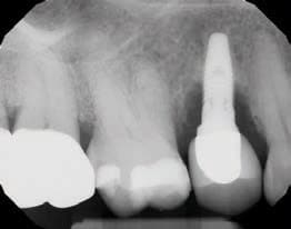

Case 7: Endoscopic debridement of retained cement from implant #12, with adjunctive irrigation of povidone-iodine (figures 9A through 9C)
Treatment by John Y. Kwan, DDS
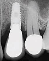
Case 8: Endoscopic debridement implant #30 with retained cement, using adjunctive systemic antibiotic amoxicillin and metronidazole, and a 5-minute soak of povidone-iodine at the time of treatment (figures 10A and 10B). Note: It should not be assumed that new bone has attached to previously diseased surfaces in these cases
Treatment by Amy Curran, RDH, in the periodontal office of Ronald Watkins, DDS, MS

key takeaways
- A 2015 systematic review and meta-analysis of nonsurgical treatment for patients with chronic periodontitis conducted by the American Dental Association Council on Scientific Affairs concluded that conventional scaling and root planing — with or without adjuncts — should be considered the initial treatment modality for patients presenting with this chronic disease.1
- The periodontal endoscope facilitates visualization of the subgingival environment via a fiber-optic probe. This system enlarges the image up to 48 times, while also delivering light and irrigation to the working field.
- Among the clinical applications for the periodontal endoscope are patients undergoing initial periodontal therapy with probing depths greater than 4 mm, and periodontal maintenance patients with chronic inflammation, or increasing probing depths.
- Additional subgingival pathologies that can be identified with the periodontal endoscope include caries, open restoration margins, cervical enamel projections, root fractures, endodontic root perforations and resorption.
- Endoscopic root debridement via hand instruments or ultrasonic inserts/tips allows for microvisual instrumentation
LIMITATIONS AND CONCERNS
Periodontal endoscopy requires a learning phase in order to become comfortable with the technology. A learning curve of approximately 10 to 30 cases is typically required to feel proficient with image interpretation, implementation of the two-handed technique, and proper patient and operator positioning (Table 2).
 Currently, training in periodontal endoscopic debridement techniques can take place in-office using online video review of the technology and instrument placement, bench training for the two-handed technique, and hands-on patient treatment. This type of instruction can be provided in private practices, but may also be implemented as part of dental, dental hygiene and periodontal clinical training.
Instruments are positioned by direct vision into the oral cavity and the operator then focuses on the monitor to govern movements. Medium to medium-plus power is used with ultrasonic instrumentation, using lateral pressure for more power (which is contrary to most teaching, but the benefit is very evident when cleaning endoscopically). More tenacious deposits require more amplitude or ultrasonic power, combined with smaller movements and more pressure over deposits until the area is completely clean.
Periodontal endoscopy utilizes a two-handed technique in which the endoscope is used in the nondominant hand (similar to holding a dental mirror), with the ultrasonic instrument in the dominant hand, moving together around the tooth during instrumentation. Subgingival visualization has shown that instrument access is far more predictable and efficient when the root surface and instrument can be viewed simultaneously. Rarely, a “view, instrument and view” technique is used when both the endoscope and ultrasonic instrument are unable to simultaneously access the area being scaled. Beginning and finishing with one explorer in each segment before starting with another explorer is an integral part of the systematic approach to endoscopic debridement. This methodology is not unlike that taught for blind, closed-pocket instrumentation.10
Ultrasonic instruments are the first choice for use with the periodontal endoscope. The most commonly used UITs are small and probe-like. Endoscopically, ultrasonic instruments provide efficient root debridement, and the technique requires a small array of UITs. A full mouth debridement typically requires only a straight, probe-like universal UIT, with an occasional need for curved or angled instruments. These nonbladed instruments are also less likely to remove healthy root structure. Efficiency is enhanced by fewer instrument changes and better instrument adaptation.10 A recent study suggests the use of hand instruments when removing cements from implants, however, due to the potential for powered instruments to eject small particles of cement into soft tissues.20 Ultimately, the choice is up to clinicians’ best judgment.
Diamond-coated ultrasonic instruments are used for advanced instrumentation in the removal of rough (globular) cementum, tenacious calculus, overhanging restorations and subgingival enamel anomalies. Because of their cutting power, however, using diamond-coated UITs requires advanced skills. This is not only true when utilizing the cutting function, it’s also necessary to avoid damage to the explorer shield, sheath or the endoscope fiber itself.10
Visualization with the dental endoscope is sometimes difficult in pockets less than 4 mm because the water clearing the camera lens is not as constant on the tooth surface as it is inside a deeper periodontal pocket. Heavy inflammation may create excessive bleeding and soft tissue tags that may float in front of the camera lens, obscuring visualization. These tissue tags often work loose as sites are debrided and “clear up,” and/or can be moved aside with the ultrasonic instrument.
CONCLUSION
Ultrasonic endoscopic periodontal debridement is a minimally invasive microvisual technology primarily utilized for the nonsurgical treatment of periodontal disease. Yet the dental endoscope also has applications for the evaluation of caries, root fractures, root resorption or perforations, residual cement around tooth and implant restorations, and subgingival anomalies. As with any dental instrumentation modality, this technology requires focused attention, training, practice and patience.
The 2015 ADA Center for Evidence-Based Dentistry clinical practice guidelines on the nonsurgical treatment of chronic periodontal disease note that scaling and root planning are still the gold standard for nonsurgical therapy.22 The periodontal endoscope supports this therapeutic approach, and has been found to be a minimally invasive technology that provides visualization with magnification and illumination within a closed pocket to aid in the diagnosis and treatment of chronic periodontitis and peri-implant disease.
References
- Smiley CJ, Tracy SL, Abt E, et al. Systematic review and meta-analysis on the nonsurgical treatment of chronic periodontitis by means of scaling and root planing with or without adjuncts. J Am Dent Assoc. 2015;146:508–524.
- Allen DL, Kerr DA. Tissue response in the guinea pig to sterile and non-sterile calculus. J Periodontol. 1965;36:121–126.
- Tan BKT, Mordan NJ, Embleton J, Pratten J, Galgut PN. Study of bacterial viability within the human supragingival dental calculus. J Periodontol. 2004;75:23–29.
- Blue CM, Lenton P, Lunos S, Poppe K, Osborn J. A pilot study comparing the outcome of scaling/root planing with and without Perioscope technology. J Dent Hyg. 2013;87:152–157.
- Rabbani GM, Ash MM, Caffesse RG. The effectiveness of subgingival scaling and root planing in calculus removal. J Periodontal. 1981;52:119–123.
- Caffesse RG, Sweeney PL, Smith BA. Scaling and root planing with and without periodontal flap surgery. J Clin Periodontol. 1986;13:205–210.
- Sherman PR, Hutchens LH Jr., Jewson LG, Moriarty JM, Greco GW, McFall WT Jr. The effectiveness of subgingival scaling and root planing. I. Clinical detection of residual calculus. J Periodontol. 1990;61:3–8.
- Stambaugh RV, Dragoo M, Smith DM, Carasali L. The limits of subgingival scaling. Int J Periodontics Restorative Dent. 1981;1:30-41.
- Perioscopy System Operators Manual, Page 6. Danville Materials, LLC, a division of Zest Anchors, LLC.
- Harrel SK, Wilson TG Jr., eds. Minimally Invasive Periodontal Therapy: Clinical Techniques and Visualization Technology. Hoboken, NJ. Wiley-Blackwell. 2015.
- Stambaugh RV, Myers G, Ebling W, Beckman B, Stambaugh K. Endoscopic visualization of the submarginal gingiva dental sulcus and tooth root surfaces. J Periodontol. 2002;73:374–382.
- Osborn JB. Role of the dental endoscope in calculus detection. Dimensions of Dental Hygiene. 2016;14(2):40,42–44.
- Wilson TG Jr., Carnio J, Schenk R, Myers G. Absence of histologic signs of chronic inflammation following closed subgingival scaling and root planning using the dental endoscope: Human biopsies — A pilot study. J Periodontol. 2008;79:2036–2041.
- Wilder RS. A new option for local delivery. Dimensions of Dental Hygiene. 2003;1(2):24–27.
- Slots J, Ting M. Systemic Antibiotics in the treatment of periodontal disease. Periodontal 2000. 2002;28:106–176.
- Slots J. Selection of antimicrobial agents in periodontal therapy. J Periodontal Res. 2002;37:389–398.
- Kotsilkov K, Emilov D, Popova CHR. Subgingival Irrigations With Povidone-Iodine as Adjunctive Treatment of Chronic Periodontitis. Journal of IMAB — Annual Proceeding (Scientific Papers). 2009;book 2. Available at: journal-imab-bg.org/statii-09/vol09_2_84-88str.pdf. Accessed July 7, 2016.
- Hoang T, Jorgensen MG, Keim RG, Pattison AM, Slots J. Povidone-iodine as a periodontal pocket disinfectant. J Periodontal Res. 2003;38:311–317.
- Wilson TG Jr. The positive relationship between excess cement and peri-implant disease: a prospective clinical endoscopic study. J Periodontol. 2009;80:1388–1392.
- Wilson TG Jr., Valderrama P, Burbano M, et al. Foreign bodies associated with peri-implantitis human biopsies. J Periodontol. 2015;86:9–15.
- Hughes K. Implant Failures Caused by Cements. Inside Dentistry. 2013;9(2). Available at: dentalaegis.com/id/2013/02/implant-failures-caused-by-cements. Accessed July 7, 2016.
- ADA Center for Evidence-Based Dentistry. Nonsurgical Treatment of Chronic Periodontitis, Clinical Practice Guideline 2015. Available at: ebd.ada.org/en/evidence/guidelines/nonsurgical-treatment-of-chronic-periodontitis. Accessed July 7, 2016.
From Decisions in Dentistry. August 2016;2(08):10–14.
