
Guest Editorial: Dental Sleep Medicine — Are You Asleep at the Wheel?
Understanding the etiology of sleep-related breathing disorders, as well as effective treatment planning and management, are pivotal to successful dental sleep medicine outcomes.
How many times have you peered into the oral cavity and noticed that your patient has massive tonsillar tissue, or a battered and red uvula, or a tongue so muscular and large that you wished your assistant had the strength of Hercules to hold it out of the way while you work?
Have you ever noticed large scalloping of the tongue on a patient and just looked past it? Or perhaps you have experienced a reaction of horror when you begin leaning the patient back in the chair and he or she insists you not lean it back any farther. Most of the time during my career, I chalked it up to “that’s just the way it is.”
These were all things I noticed in my practice and never thought much about until I started investigating the possible causalities of these findings. What all these scenarios have in common is they are potential signs of a sleep-related breathing disorder (SRBD).
According to the American Academy of Sleep Medicine, SRBDs are “sleep disorders that involve difficulty breathing during sleep.” The most common is obstructive sleep apnea (OSA). These disorders can present as excessive daytime sleepiness, insomnia, sleep-related eating disorders, snoring, bruxism, sleep leg cramps, and restless leg syndrome — to name just a few. It is estimated that approximately 70 million people experience sleep disorders each year.1
SLEEP DISORDER DIAGNOSIS
A sleep disorder is a medical condition that is diagnosed by a sleep physician who is specifically trained in sleep medicine. Typically, pulmonologists, otolaryngologists and neurologists will have additional training in this specialty. In addition, providers specializing in internal medicine, cardiology, psychiatry, pediatrics or anesthesiology often pursue residencies and fellowships that can lead to a sleep medicine subspecialty. These individuals often attain board certification in sleep medicine.
What does this have to do with dentistry? I once asked that question myself until I began investigating the challenges I saw presenting every day in practice. Patients or their significant others would come in complaining about their partner’s snoring and wanting to know if I could help before they did something rash. Many were sleeping in separate bedrooms because they could not tolerate the other person’s snoring. I figured I could help and started creating snoring devices. I was a “hero” and it felt good. That is, until the devices broke or the teeth began to shift. I stopped that practice because I did not want to play orthodontist and put teeth back where they were originally located.
Then articles began popping up in various journals about the creation of mandibular advancement devices that could help patients with OSA. Again, I thought that would be easy — take a couple of impressions, send it off to the lab, get it back, and simply adjust the device until the patient reports that he or she is no longer snoring.
GETTING THE BAND TOGETHER
Some of the devices had polymer-elastic bands that would hold the jaw forward and offer patients the ability to move their jaw sideways as needed. I explained to the physicians I was working with that this would be a great help to their patients.
Then the bands connecting the upper and lower portions began breaking on some devices, and too often patients’ snoring was not mitigated as the devices were advanced forward. And again, teeth began to move and joints began to hurt. To make matters worse, the secondary sleep studies that patients had — as required by their physician — did not show improvement in the outcome. What did I get myself into?
What seemed so simple became a nightmare. I started investigating different types of devices that claimed to be the solution to these problems. But, alas, there was no improvement. Then the sleep physician that I worked with called to say she could not send any more patients to me because the secondary polysomnograms did not show improvement. To put it bluntly, I had “egg on my face.”
SCIENCE OF DENTAL SLEEP MEDICINE
As I investigated further into the science of dental sleep medicine, I learned the methodology I was using was not an exact science, and that it could not provide consistent outcomes, as I presumed it would. The George Gauge (GG) method I was using to create the plastic devices is very explicit in the accompanying instructions. The passage “Where Is the Correct Construction Bite”2 states: “The optimum position of the construction bite varies in each patient. It obviously is somewhere between centric relation and full protrusive. There are no landmarks that can be used to accurately locate it. No relationship of upper-to-lower incisors can be correct for all patients.”
Understandably, some authors have suggested there are inherent challenges with this methodology. In their paper, “Retrospective comparison of the George Gauge registration and sibilant phoneme registration for constructing OSA oral appliances,” Viviano et al3 state, “The George Gauge registration (GGR) has become a standard method used to document mandibular advancement … Notwithstanding that GGR overestimates measurements of advancement when compared to a ruler and cephalometric measurements.” The authors further note, “The literature regarding optimum advancement is also conflicted, some determining optimum benefit with 70% advancement and others finding no benefit beyond 50% advancement.”3
Hamoda et al4 suggest the long-term effects of oral appliance therapy are maxillary incisor retroclination and mandibular proclination, and that changes appear to continuously progress with time. Hu and Comisi5 demonstrated the position determined with the 70% George Gauge was, on average, 5.0 mm more protrusive than positions found using an alternative position-determining device called a pharyngometer.
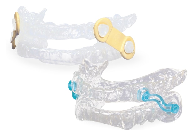
Pharyngometry is an analysis tool used in the determination of optimal treatment position of an oral appliance when managing OSA. It aids in identifying the location and severity of pharyngeal obstruction via an analysis of the minimal cross-sectional area. This minimally invasive testing mechanism can help screen patients suspected of having OSA — and, after a diagnosis is rendered by a sleep physician, it can also help determine the optimal jaw position for appliance therapy (Figure 1). Furthermore, it can be a major asset in determining, prior to the creation of an oral device, if the patient is likely to respond to an appliance.
This understanding of whether a patient will respond to therapy is critical since all too often with other approaches there is no way of knowing if the device will be effective until the patient wears it. This, I came to realize, is where the problems developed in my dental sleep medicine practice. I was creating devices without knowing what the end results would be. I was hopeful for success, but consistent success — at least for me — proved elusive.
A NEW APPROACH
At the insistence of my spouse, Karen Comisi, we attended a course on a new way to approach dental sleep medicine. It included rhinometry and pharyngometry. From that moment on, everything changed. I realized this was the missing piece to the puzzle of treating patients with SRBDs. I returned to my sleep physician colleagues with this new concept, presented the information, and requested they provide just two patients for me to treat with this technology. If I failed, I would not darken their doors again. If I succeeded, then I requested they send more patients for me to help manage.
Pharyngometry employs acoustic sound waves that travel down the airway of the patient in a noninvasive manner to help identify where the problem in the airway might be and determine the most beneficial and therapeutic position for an oral device. This typically is found by utilizing special airway jigs that can help determine therapeutic position by opening the vertical bite at 4, 6, 8, 10 and 12 mm while using the pharyngometer. Typically, most devices I create are at an edge-to-edge position at this vertical dimension. If advancement is needed, these jigs can also help place the jaws in those positions while working with the pharyngometer. Ultimately, this process places less stress on the temporomandibular joint area and enhances patient compliance.
The first two patients I treated using this new methodology responded well. Both were happy with the results, as indicated by great secondary sleep studies. In turn, their treating physicians were happy, and, of course, so was I. The floodgates opened, and my dental sleep medicine practice grew. The literature is currently filled with evidence of the benefits of incorporating this methodology when creating devices for patients who have been referred for an oral appliance.6–11
ENHANCED SCREENING
This marked another watershed moment in my dental sleep medicine journey. I began to learn about the oral signs of SRBDs and started incorporating enhanced screening. We discovered the bruxism we were attempting to treat could often be traced to sleep issues, and there was a possibility that placing mouth guards to protect the teeth could be closing off the airway.12 Now, when I examine a patient with obvious bruxing, I always inquire about sleep issues. This requires clinicians to become real investigators — but it is worth it. Even more has been published on this topic.13–16
If the examination and updated patient record affirm a history of sleep issues, diagnosis of OSA, snoring or excessive daytime sleepiness, a single-arch, flat-plane splint is not going to be created. Typically, I will refer the patient to a sleep medicine specialist and request an evaluation and a sleep study to render a diagnosis. If the patient is diagnosed with OSA and a prescription for an oral appliance is provided, I will then move forward with the fabrication of a custom therapeutic device using pharyngometry.
IN SUMMARY
It is important to remember SRBDs are medical diseases and a prescription from a sleep physician is needed to provide dental sleep medicine care for these patients — in fact, all patients with OSA. The days of making flat-plane mouth guards and blindly creating sleep appliances are gone from my practice and from what I now teach as part of the sleep medicine curriculum at the Medical University of South Carolina.
The physicians I work with here and the students I teach have a greater understanding of the partnership between dentistry and medicine in this area. Our students are gaining further insights regarding the need to screen all patients for potential SRBDs. In this way, more people will be screened and ultimately treated for this ubiquitous medical disease.
As oral health professionals, we are on the front lines. Now it’s your turn to learn more about dental sleep medicine, the underlying etiologies, and how you can help the patients who you did not previously recognize as presenting with sleep disorder issues.
Key Takeaways
- Massive tonsillar tissue, a battered and red uvula, or a muscular, unusually large or scalloped tongue are potential signs of sleep-related breathing disorders (SRBDs), such as obstructive sleep apnea (OSA).
- These disorders can present as excessive daytime sleepiness, insomnia, snoring, bruxism, sleep leg cramps, and restless leg syndrome — to name just a few.
- Rhinometry and pharyngometry are key concepts for dental providers who treat patients with SRBDs.
- Pharyngometry can help determine the optimal treatment position of an oral appliance when managing OSA.
- Additionally, this minimally invasive testing mechanism can be used to screen patients suspected of having OSA — potentially leading to a referral to a sleep medicine physician for diagnosis.
- Furthermore, pharyngometry can be a major asset in determining, prior to the creation of an oral appliance, if the patient is likely to respond to this type of treatment.
- It is important to remember SRBDs are medical diseases and a diagnosis and prescription from a sleep physician are required before providing dental sleep medicine treatment.
REFERENCES
- American Academy of Sleep Medicine. Sleep Medicine. Available at: https://sleepeducation.org/sleep-disorders/. Accessed November 10, 2022.
- Great Lakes Dental Technologies. The George Gauge. Available at: https://www.greatlakesdentaltech.com/media/resources/GeorgeGaugeInstructions.pdf. Accessed November 10, 2022.
- Viviano J, Klauer D, Olmos S, Viviano JD. Retrospective comparison of the George Gauge registration and the sibilant phoneme registration for constructing OSA oral appliances. Cranio. 2022;40:5–13.
- Hamoda MM, Almeida FR, Pliska BT. Long-term side effects of sleep apnea treatment with oral appliances: nature, magnitude and predictors of long-term changes. Sleep Med. 2019;56:184–191.
- Hu JC, Comisi JC. Vertical dimension in dental sleep medicine oral appliance therapy. Gen Dent. 2020;68:69–76.
- DeYoung PN, Bakker JP, Sands SA, et al. Acoustic pharyngometry measurement of minimal cross-sectional airway area is a significant independent predictor of moderate-to-severe obstructive sleep apnea. J Clin Sleep Med. 2013;9:1161–1164.
- Kim BY, Cho JH, Kim DH, et al. Utility of acoustic pharyngometry for screening of obstructive sleep apnea. Auris Nasus Larynx. 2020;47:435–442.
- Kamal I. Test-retest validity of acoustic pharyngometry measurements. Otolaryngol Head Neck Surg. 2004;130:223–228.
- Rong T, Ma JG, Li XY, Cai L, Jiang XX, Wang YX. The application of acoustic pharyngometry in diagnosis and treatment of OSAHS. J Clin Otorhinolaryngology Head Neck Surg. 2018;32:477–480.
- Zhao Y, Li X, Ma J. Combined application of pharyngeal volume and minimal cross-sectional area may be helpful in screening persons suspected of obstructive sleep apnea (OSA). Sleep Breath. 2022;26:243–250.
- Friedman M, Samuelson CG, Hamilton CS. Acoustic pharyngometry: a substitute for drug-induced sleep endoscopy? Otolaryngol Head Neck Surg. 2012;147(Suppl 2):P119.
- Gagnon Y, Mayer P, Morisson F, Rompré PH, Lavigne GJ. Aggravation of respiratory disturbances by the use of an occlusal splint in apneic patients: a pilot study. Int J Prosthodont. 2004;17:447–453.
- Martynowicz H, Gac P, Brzecka A, et al. The relationship between sleep bruxism and obstructive sleep apnea based on polysomnographic findings. J Clin Med. 2019;8:1653.
- Oksenberg A, Arons E. Sleep bruxism related to obstructive sleep apnea: the effect of continuous positive airway pressure. Sleep Med. 2002;3:513–515.
- Balasubramaniam R, Klasser GD, Cistulli PA, Lavigne GJ. The link between sleep bruxism, sleep disordered breathing and temporomandibular disorders: an evidence-based review. J Dent Sleep Med. 2014;1:27–37.
- Mayer P, Heinzer R, Lavigne G. Sleep bruxism in respiratory medicine practice. Chest. 2016;149:262–271.
From Decisions in Dentistry. December 2022;8(12)18-19,21-22.
