

Cosmetic Considerations for the Single Anterior Implant Restoration
Mastering the art of these restorations requires meticulous planning, precise execution, and a deep understanding of esthetic principles.
Restoring a single central incisor is one of the most challenging procedures in dentistry. In the case of a single central implant restoration, challenges may become twofold. Osseointegration of a single anterior implant does not equate to a successful implant; esthetic results are among the critical criteria that must be considered for success.1 Dentistry is an art, and dentists have the unique responsibility of improving or maintaining function while also enhancing and empowering a smile. Fabricating a chairside provisional implant crown with the goal of achieving a natural emergence profile and gingival contour can be instrumental in achieving a more ideal final result.
Several criteria must be considered when designing the ideal smile. The lips are considered the frame of the smile, and are one factor in the amount of gingival display during function. Further is the position of the gingival zenith.
According to Pawar et al,2 the gingival zenith is defined as the most apical point of the gingival marginal scallop. This critical landmark has a specific ideal orientation in the apicocoronal and mesiodistal directions. The gingival line is defined as the line joining the tangents of the gingival zeniths of the central incisor and canine. The gingival zenith of the canine is ideally apical to the zenith of the central incisor, and the gingival zenith of the lateral incisor is below that gingival line.2 Additionally, the gingival zenith of the central incisor should be at the distal third of the tooth, which helps determine the appropriate long axis of the tooth and line angle position.3
The “golden proportions” with regard to the anterior dentition may provide an objective starting point for a balanced smile, although they should be considered a guideline not a rule.4 Research shows the average length of the maxillary central incisor is 10.0 to 10.5 mm, with an ideal height-to-width ratio of 1.2 to 1. The canines are generally similar in length to the central incisors, while the lateral incisors are shorter at 8.2 mm on average.5
While the golden proportions certainly provide a useful guide, artistic license is necessary for a natural appearance, especially in the maxillary anterior. Some asymmetry and variation from the norm are present in most smiles, and these must be harnessed to create a natural, life-like prosthesis.
Case Study: Initial Evaluation
A 32-year-old woman presented with a previously restored #9 implant with the chief complaint that the implant does not resemble a natural tooth. To determine why the implant crown stood out, a comprehensive esthetic diagnosis was required. One way to do this is to begin with the end result in mind, and work backward. This method entails beginning the case with a clear vision of the desired outcome, and then taking proactive steps to accomplish the defined goal. In this case, the desired outcome for the patient was a balanced smile in which her implant crown did not stand out.
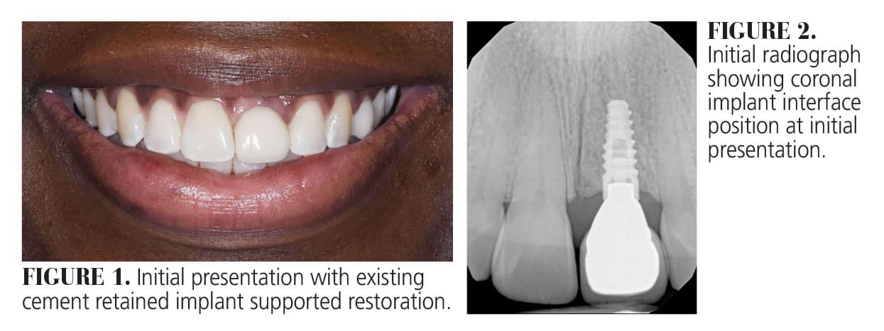
To determine a plan of action, esthetic criteria, including smile line, lip posture, gingival display, incisal edge position, golden proportions of the anterior dentition, tooth color, tooth shape, and gingival zenith, were used to determine what needed to be altered. This particular patient presented with an appropriate smile frame and some gingival display (0.5 to 3 mm) based on her lip position while smiling. This is considered ideal for a young woman (Figure 1).6 Further, the incisal edges of her maxillary teeth follow the natural curve of her lower lip, and the length of her natural incisor #8 was 10.5 mm, considered ideal according to the golden proportions. The color of the existing implant crown was generally harmonious with the surrounding dentition.
The shape of the central incisors were asymmetrical; the right central incisor appeared triangular, while the shape of her left central incisor appeared round. Most important was the gingival zenith of the left central incisor, which presented below the gingival line and within the mesial third of the tooth. This negatively impacted the visualization of the long axis of the tooth, especially when contrasted with the adjacent central incisor. It created a cant in the midline, and drew the eye to the implant crown. Because of the gingival zenith position of #9, the harmony of the entire smile was disrupted. Therefore, it was determined that both the gingival zenith of #9 and the shape of the central incisors would have to be altered in hopes of creating a more natural restoration.
Implant Limitations
Implants should be surgically placed with an ideal prosthesis planned. Screw-retained restorations are often considered preferable over cement-retained restorations because they reduce the risk of cement reaching the abutment interface. This also increases ease of access if needed. Implants should be placed apical to the cementoenamel junction (CEJ) of the adjacent tooth if possible for a more ideal emergence design with a custom abutment.7
Figure 2 shows the implant had been placed in a relatively coronal position, approximately even with the adjacent central incisor CEJ, limiting the space to work with the emergence profile of the implant crown and custom abutment. In this case, beginning with a provisional implant crown for tissue training was primarily diagnostic in nature to see if a more ideal gingival zenith could be accomplished.
Clearly defined parameters have been set to achieve ideal tissue height and papilla fill. Numerous studies have shown that the interproximal contact point between adjacent teeth should be 5 mm from the crest of bone to allow papilla fill. At 6 mm, the chance of papilla fill drops to 50%.8,9 To achieve a more ideal contact point and shape of adjacent central incisor, a matrix system was utilized in a conservative additive composite bonding technique to change the mesial contour of #8- changing the shape from triangular to ovoid and allowing an ideal interproximal contact 5 mm from the crest of bone to allow papilla fill, effectively avoiding the dreaded “black triangle.”
Chairside Provisional Technique
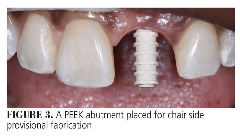
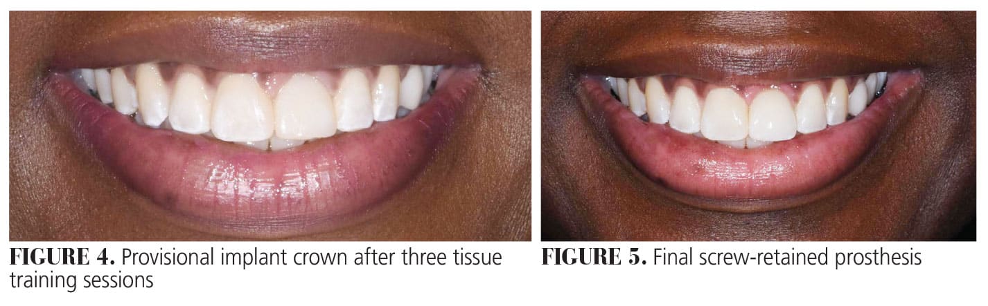
To alter the gingival zenith of #9, a tissue training technique using a chairside provisional implant crown was utilized to gently guide the tissues into the ideal position. A PEEK abutment was seated on the implant, and a matrix applied to fabricate a chairside provisional (Figure 3). The provisional crown was modified at the gingival aspect to idealize emergence. Composite resin was added to the facial, blanching the tissue and gently pushing it into a more apical position. For this case, the patient returned every 4 to 6 weeks for three sessions to gradually apply more composite at the facial gingival emergence profile until the desired gingival zenith was reached (Figure 4).
 This technique can also be used as an immediate provisional technique if primary stability is achieved at the time of implant placement, so the tissue management begins the day of implant surgery.10 The provisional crown can then be modified and the gingival zenith position perfected. While more time consuming than a laser or surgically removing excess gingival tissue, this technique gently pushes up the keratinized tissue to maintain a level mucogingival junction (MGJ) rather than sacrificing healthy keratinized tissue and creating a discrepancy in the MGJ with adjacent teeth.
Once the tissue is positioned appropriately, photos and an impression or digital scan of the provisional implant crown can be sent to the lab to replicate the gingival emergence profile of the implant crown. This can be achieved with a digital impression or intraoral scan with a scan body immediately after provisional crown removal to provide a digital model of the site to the lab. With an analog impression, tissue collapse during the impression can be avoided by placing flowable composite around the impression coping immediately after the removal of the provisional prosthesis.
Final Prosthesis
Figure 5 shows the final seated prosthesis. This is a screw retained zirconia implant crown with a custom gold hue titanium abutment. Zirconia was selected for its strength and cosmetic benefits, but also for its opaque nature. The gingival zenith is more apical and distal when compared to the original prosthesis shown in Figure 1.
The midline cant was corrected by changing the shape of the adjacent central incisor with additive composite bonding. When applying objective esthetic criteria to the patient’s smile after the prosthesis replacement, ideal criteria are met, resulting in a pleasing, harmonious, balanced smile. Managing this case with a provisional crown first and transferring the emergence profile to the lab made it more predictable for both the laboratory technician and clinician. The more information provided to the lab, the better.
To empower a smile can mean to create harmony in which a tooth does not stand out, but rather the maxillary anterior teeth function together to provide a pleasing balance in function and esthetics. This can be an especially challenging feat when restoring a single anterior implant. This balance can be accomplished by utilizing esthetic criteria for a more ideal form and function, and planning for such a result from the time of surgical placement.
Acknowledgment
The author would like to thank Jeremy Pearson, owner and laboratory technician at Exquisite Smile Designs in Eddy, Texas, for his beautiful work on this case.
References
- Smith DE, Zarb GA . Criteria for success of osseointegrated endosseous implants. J Prosthet Dent. 1989;62:567–572.
- Pawar B, Mishra P, Banga P, Marawar PP. Gingival zenith and its role in redefining esthetics: a clinical study. J Indian Soc Periodontol. 2011;15:135-138.
- Martins FV, Mattos de Santana CM, Magno MB, Maia LC, Fonseca EM, Barcellos de Santana R. Gingival zenith level, position, and symmetry: A systematic review and meta-analysis. J Prosthet Dent. 2024:S0022-3913(24)00225-7.
- Akl MA, Mansour DE, Mays K, Wee AG. Mathematical Tooth Proportions: A Systematic Review. J Prosthodont. 2022;31:289-298.
- Robbins MA, William J, Rouse JS. Chapter 2: global analysis diagnosis form: length of maxillary anterior teeth. Global Diagnosis: A New Vision of Dental Diagnosis and Treatment Planning. Hanover Park, Illinois: Quintessence Publishing Co; 2016:21–22.
- Mahn DH, Reangber S. Gingival display in “forced” smile evaluated by sex and age. Compend Contin Educ Dent. 2022;43:e9-e12.
- Buser D, Martin W, Belser UC. Optimizing esthetics for implant restorations in the anterior maxilla: anatomic and surgical considerations. Int J Oral Maxillofac Implants. 2004:19Suppl:43-61.
- Choquet V, Hermans M, Adriaenssens P, et al. Clinical and radiographic evaluation of the papilla level adjacent to single-tooth dental implants. A retrospective study in the maxillary anterior region. J Periodontol. 2001;72:1364–1371.
- Luo R, Zhu Z, Huang J, Ye Y. The esthetic outcome of interproximal papilla between implant-restored unilateral and bilateral maxillary central incisors: a cross-sectional comparative study. Int J Oral Maxillofac Implants. 2022;37:1063-1070.
- Qin R, Chen Y, Han C, Wu D, Yu F, He D. Immediate implant placement with or without immediate provisionalization in the maxillary esthetic zone: a systematic review and meta-analysis. Int J Oral Maxillofac Implants. 2023;38:422-434. 1. Smith DE, Zarb GA . Criteria for success of osseointegrated endosseous implants. J Prosthet Dent. 1989;62:567–572.
From Decisions in Dentistry. January/February 2025;11(1):18-21.
