
 TRILOKS/ISTOCK/GETTY IMAGES PLUS
Clinical Signs and Risk Factors for Autoimmune Diseases
Oral health professionals can play a vital role in ensuring that patients who present with signs or risk factors for these serious conditions receive an appropriate referral.
PART 1 of a two-part series. The first article in this two-part series explores the clinical signs and risk factors for autoimmune diseases. Appearing in a future issue, Part 2 will cover the medications used to treat autoimmune diseases.
PURCHASE COURSE
This course was published in the December 2021 issue and expires December 2024. The author has no commercial conflicts of interest to disclose. This 2 credit hour self-study activity is electronically mediated.
This course was published in the December 2021 issue and expires December 2024. The author has no commercial conflicts of interest to disclose. This 2 credit hour self-study activity is electronically mediated.
EDUCATIONAL OBJECTIVES
After reading this course, the participant should be able to:
- Explain the prevalence of autoimmune diseases (ADs).
- Discuss AD risk factors.
- Identify ADs with manifestations identifiable by oral health professionals.
Autoimmune diseases (AD) are a complex and heterogeneous group of more than 100 recognized conditions caused by immune system dysfunction.1–3 As ADs impact approximately one in five Americans, oral health professionals will likely encounter patients with undiagnosed conditions. Consequently, dental teams should be prepared to recognize their clinical manifestations and refer patients for early medical care.2–5
The immune system involves the interaction of T-lymphocyte cells (T-cells), B-lymphocyte cells (B-cells), and other immune cells that protect the host by subduing the effects of foreign substances. To prevent immune cell overproduction and attack of host cells, the body uses three mechanisms — clonal deletion, clonal anergy, and suppression of autoreactive lymphocytes — to remove or suppress autoreactive T- and B-cells.2,6–10 T-regulator cells (Tregs) protect against excessive autoreactivity, but inflammatory environments can create an imbalance by stimulating effector T-cells and diminishing Tregs. This imbalance increases immune cell production, initiates tissue damage, and is a contributing factor in many ADs.6,11,12 In patients with ADs, the immune system ceases to differentiate between host cells and foreign antigens, and consequently targets both.3 All ADs have the ability to cause chronic and severe tissue damage.2–4,8,13,14
The American Autoimmune-Related Disease Association has identified more than 100 ADs, affecting approximately 20% of the U.S. population.3,4 Chronic, inflammatory and rheumatic diseases comprise the largest group of ADs; with rheumatoid arthritis in adults and juvenile idiopathic arthritis in children accounting for the largest number of cases.15 In general, ADs are more prevalent among women, comprising 80% of the diagnoses.2,3,13,16 Among young to middle-aged women, ADs are among the leading causes of morbidity and mortality.13,17
RISK FACTORS FOR AUTOIMMUNE DISEASES
Many ADs have identifiable risk factors, but are not associated with one cause. Nonmodifiable risk factors include genetics, hormones and infections, while modifiable risk factors encompass environmental exposures, medications and lifestyle.2,18 Oral health professionals may help susceptible patients identify healthy behavioral modifications to reduce their AD risk.19
These disorders are typically multigenic, with many contributing genes.6,12 The overexpression, suppression or alteration of genes involved with immune cells increase AD risk. Specific alleles, alternative forms of genes, and human leukocyte antigen molecules are also associated with ADs. Rather than a specific genetic alteration or number of susceptible genes, the main determining factor in disease development is host genetic susceptibility outweighing the counterbalance of protective genes.6,12
The quantity of X chromosome immunity genes is a possible reason for a strong female predominance in AD — two Xs double the mutation risk.3,16,20 One theory suggests that inadequate suppression of X immunity genes causes an overproduction of autoreactive T-cells. Another hypothesis proposes that X inactivation generates a genetically favored X; cells with the less favored X may trigger autoimmunity.3
Hormone fluctuations or medications can impact the development, initiation and severity of AD in women.3,4,20 Increased estrogen associated with puberty, pregnancy or medication use is anti-inflammatory, but amplifies systemic sclerosis and systemic lupus erythematosus risk, while progesterone-only oral contraceptives are protective against systemic lupus erythematosus.2,3,19 Estrogen reduction with menopause increases inflammatory AD risk. Infections are also associated with autoimmunity and ADs.4,12,14 Pathogens can prompt molecular mimicry and bystander activation, or stimulate autoreactive lymphocytes or reactivate antigens. Subsequent infections can trigger a cytokine storm.8
Nearly 100 medications are associated with AD development,9,16 with drug-induced lupus being the most common. Pulmonary, cardiovascular, hepatic, and other systemic impacts can also occur, resulting in a systemic lupus erythematosus diagnosis; 10% or more of systemic lupus erythematosus cases initially develop drug-induced lupus.9 When possible, eliminating or changing the medication upon early stage drug-induced lupus recognition may reduce symptoms within weeks, but autoantibodies can remain up to two years.9
High levels and duration of chemical or environmental pollutant exposures can trigger autoreactivity and subsequent AD.6,16 Air pollution particulates and exposures to asbestos, crystalline silica, mercury and solvents are associated with ADs. Extended childhood exposure to agricultural pesticides and insecticides is associated with systemic lupus erythematosus and rheumatoid arthritis.16
Lifestyle choices can impact the risk of developing ADs. Obesity increases systemic inflammation, while a calorie restriction diet can reduce inflammatory cytokines and AD symptom severity.16 Vitamin D deficiency is related to increased AD risk.8 Nutritional counseling and a balanced, healthy diet may reduce disease risk. Psychological and environmental stressors can alter immune system responses. Long-term stress and similar conditions, such as post-traumatic stress disorder or acute stress reaction, reduce cortisol levels, increasing the risk of immune dysfunction and AD. Patients who avoid treatment or individuals with a psychiatric comorbidities are at risk for ADs.21 Encouraging patients to seek medical and psychological support is an important recommendation from dental team members.
Risk for AD development and exacerbation increases with cigarette smoking in both men and women.14,16,19,22 The toxins in cigarettes can stimulate autoreactive cells or inflammatory cytokines and damage DNA, leading to genetic mutation.19 Initiating tobacco cessation education and referring for additional support are key in reducing disease risk.
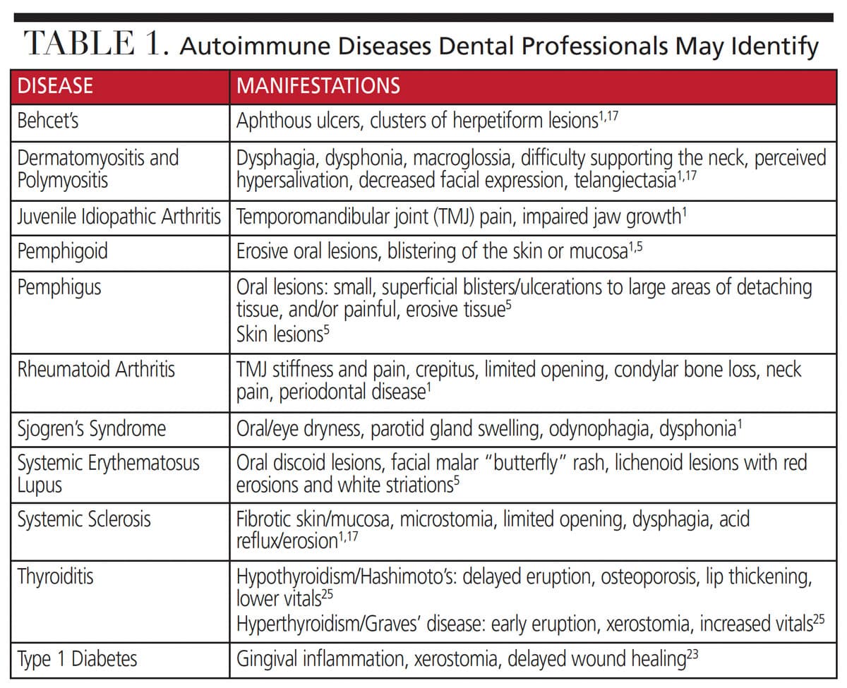
These conditions are classified into two general categories: single-organ and systemic. Oral manifestations are common initial signs of immune suppression or dysfunction that may indicate the need for medical referral.1,4,15 The following is an overview of ADs and their manifestations identifiable by oral health professionals (Table 1).
TRILOKS/ISTOCK/GETTY IMAGES PLUS
SINGLE-ORGAN AUTOIMMUNE DISEASES
Type 1 Diabetes — A single-organ AD, type 1 diabetes is caused by the destruction of insulin-secreting pancreatic B-cell by autoreactive T-cells. Risk factors include genetic susceptibility, vitamin D deficiency and viral infections, such as enteroviruses, rotavirus, congenital rubella syndrome, mumps and cytomegalovirus.8,23 Periodontal disease and diabetes negatively impact each other. Xerostomia is also common among individuals with diabetes and can result in burning sensations, caries and candidiasis.24
Thyroid Diseases — Individuals with Hashimoto thyroiditis and Graves’ disease display autoantibodies against thyroid tissue. Hashimoto thyroiditis results in hypothyroidism and damage to the thyroid follicles, while Graves’ disease causes an extensive release of thyroid hormone (hyperthyroidism).8 The cardiovascular system and metabolic rate are impacted, resulting in lower pulse rate, lethargy and weight gain with hypothyroidism, and hypertension, tachycardia and emotional instability with hyperthyroidism.25 Childhood hypothyroidism can cause macroglossia, dysguesia, lip thickening, and delayed eruption of permanent teeth, while hyperthyroidism may cause early eruption. Adult hypothyroidism may manifest as poor periodontal health, delayed wound healing and lower vitals, while hyperthyroidism may cause dry mouth, burning mouth syndrome, osteoporosis, enlarged palpable thyroid gland, and increased vital signs.25
Inflammatory Bowel Disease — This group of chronic conditions results in gastrointestinal tissue damage, and a patient’s diagnosis may include Crohn’s disease, celiac disease or ulcerative colitis. Inflammatory bowel disease may be caused by a genetic alteration resulting in exaggerated immune response to the commensal gut bacteria, rather than an autoimmune response targeting the tissue.6,12 Aphthous ulcers are common among those with inflammatory bowel disease and they are cause for medical referral if seen with gastrointestinal symptoms.15
Pemphigus Vulgaris — This AD occurs when antibodies develop against the desmosomal cells that construct skin and mucosal tissue. Its impact on adhesion causes blistering and sloughing of tissue, resulting in erosive ulcerations.5 Oral lesions are the first signs in more than 50% of patients with pemphigus vulgaris, followed by skin lesions. The oral lesions range from small, superficial blisters or ulcerations to large areas of tissue that detach with minimal pressure, exposing painful, burning erosions. These lesions may be confused with lichen planus, candidiasis and pemphigoid.5
Mucous Membrane Pemphigoid — A blistering condition developed in response to antibodies against the mucosal and epithelial basal membranes, mucous membrane pemphigoid causes detachment of the epithelium from the connective tissue.5 Intraoral and pharyngeal pemphigoid blisters are less fragile than in pemphigus, but nearly 94% also experience erosive, gingival ulcerations known as desquamative gingivitis. Pemphigoid frequently occurs in conjunction with periodontal disease, and pain, bleeding or burning sensations during mastication can also occur.5,15
SYSTEMIC DISEASES
Systemic Lupus Erythematosus — A chronic, systemic, inflammatory condition, systemic lupus erythematosus is caused by the multiple autoantibodies that develop due to the alteration of B-cells reactivity.6 Skin and oral mucosa are often damaged first, then the disease can progress to the kidneys, lungs, eyes, nervous system, joints and muscles. Organ failure is the most severe potential impact.5,8 A hormone-related disorder, it primarily affects women during puberty and the childbearing years.1,5,8,19 The risk for systemic lupus erythematosus increases with smoking, family history, estrogen use, Epstein-Barr virus exposure, and African American or Native American heritage.1,8,16,19
Early signs of systemic lupus erythematosus include the malar “butterfly” rash across the nose and cheeks (presents in 85% of affected individuals) and oral lesions of variable size, color, location and appearance.1,5 Multiple aphthous ulcers, with some chronic occurrences, are seen in up to 50% of affected patients.15 Lichenoid mucositis and discoid lesions on the lips or mucosa may resemble lichen planus, erythema multiforme or vesiculobullous lesions.1,5,15 The lichenoid lesions appear as red, erosive or ulcerated tissue with white striations, similar to lichen planus, but without symmetry.5 Up to 70% of these patients also experience periodontal disease.15
Rheumatoid Arthritis/Juvenile Idiopathic Arthritis — These chronic, inflammatory conditions instigate bilateral bone, cartilage and joint destruction.1,8,26 Pro-inflammatory cytokines and their mediators cause swelling, pain, deformity, and decreased function starting in the small joints of the extremities. Autoantibodies perpetuate the inflammation and damage.1,26 Rheumatoid arthritis risk factors include smoking and infections from the oral pathogen Porphyromonas gingivalis and the intestinal pathogen Escherichia coli.8
Temporomandibular joint (TMJ) pain and stiffness are the most common oral manifestations seen in rheumatoid arthritis/juvenile idiopathic arthritis. Nearly 90% of those with juvenile idiopathic arthritis experience TMJ involvement and it may be the only joint affected.1,15 Earlier stages manifest as swelling and discomfort during TMJ function, but, as rheumatoid arthritis/juvenile idiopathic arthritis progresses, profound TMJ pain, crepitus, limited opening and neck pain can occur. If left untreated, juvenile idiopathic arthritis can impede jaw growth or lead to jaw deviation, ankyloses and deformities.1,15
The rates of periodontal disease in patients with rheumatoid arthritis are twice that of the general population, and, as with diabetes, the severity of one condition negatively impacts the other. Periodontitis and rheumatoid arthritis share pathophysiology and risk factors, as well as an inflammatory response to oral bacteria and smoking.1,15 Xerostomia is also prevalent, with one-third of those with rheumatoid arthritis/juvenile idiopathic arthritis developing Sjörgen’s syndrome as a secondary condition.1
Sjörgen’s Syndrome — A B-cell mediated, chronic inflammatory condition, Sjörgen’s syndrome primarily affects the salivary and lacrimal glands of menopausal women. Half of all cases are primary, with the other half occurring secondary to another AD.1,5 Although xerostomia and xerophthalmia are the principal manifestations of Sjörgen’s syndrome, systemic effects on the exocrine glands can impact vascular, neurological, hepatic, respiratory and cutaneous system functions.1 Initial signs of Sjörgen’s syndrome include enlarged parotid salivary glands and hyposalivation. Patients may have adequate salivary flow during salivary tests, but have perceived xerostomia, odynophagia or dysphonia. The dryness may progress to altered taste, dysphagia, dysarthria, dysguesia, gingivitis, gingival fragility, candidiasis and caries.1,5,15
Multiple Sclerosis — This T-cell mediated disease involves central nervous system (CNS) nerve demyelination and occurs most frequently in women and white individuals.8,17,20 Sclerotic CNS tissue and subsequent nerve degeneration lead to a loss of sensation and motor, autonomic and neurocognitive function. The slowly progressing, relapsing-remitting type of multiple sclerosis is the most common and often manifests in young adulthood.20 Two events are theorized to trigger multiple sclerosis, CNS inflammation or infection resulting in fluid accumulation and cytokine migration to the area, then a second pathogen that causes nonspecific cytokines to activate autoreactive T-cells.8 Oral or facial paresthesia, neuropathy, palsy, or pain associated with the trigeminal nerve may be the first signs of multiple sclerosis. Trigeminal neuralgia can be bilateral and elicit pain at the slightest touch.17
Psoriasis — This skin disorder affects 2% to 3% of the U.S. population, regardless of age. Approximately 30% of these patients will also develop psoriatic arthritis, which destroys joints, cartilage and bone through osteoclastic activity.11 The inflammatory cascade caused by psoriasis results in the development of tumor necrosis factor alpha and additional cells, which perpetuate inflammation and tissue damage.11 Risk factors include genetic, environmental and infectious factors, including streptococcal exposure. Overexpression of an antimicrobial peptide, activation of T-cells and pro-inflammatory cytokines in the skin, and physical or psychological stresses, are linked to outbreaks. More than 90% of patients with psoriasis have plaque psoriasis, which manifests as painful or itchy raised, red scaly patches. Joint inflammation and pain occur with psoriatic arthritis, but, unlike rheumatoid arthritis, affected joints can be unilateral and localized.11
Behcet’s Disease — A condition of systemic vasculitis, Behcet’s disease affects the skin, genitals and eyes as a result of antimucous antibodies.5,15 It affects both genders in their 30s and is most prevalent among Mediterranean and Asian populations.5 Minor and major aphthous ulcers or herpetiform clusters are the first sign of disease.1,5,15 These lesions are indistinguishable from aphthous ulcers, but are seen cyclically.
Systemic Sclerosis — This systemic condition causes vascular abnormalities and fibrotic tissue in the skin and internal organs, and organ damage can occur even in early stages.1,15 Oral health professionals may recognize telangiectasias, visibly dilated blood vessels, and mucosa or skin thickening. As collagen and fibrotic tissue are deposited, a smooth mask-like appearance occurs and the size of the oral cavity is reduced. Microstomia is the most common oral manifestation leading to limited opening in more than 70% of those affected.1,15 Increased risk for tooth loss, tongue cancer or esophageal involvement (such as acid reflux or dysphagia) are also observed. Radiographically, a widened periodontal ligament and TMJ resorption may be noted.15
Myopathies — In polymyositis, the collagen tissue is affected, while dermatomyositis involves a vasculopathy of small vessels that primarily affect the skin and muscles.1 As with systemic sclerosis, mucosal telangiectasia may be seen with dermatomyositis. As the oral musculature is affected, dysphagia, dysphonia, and decreased facial expressions may become apparent. Patients may complain of hypersalivation, but this is likely related to the impaired swallowing reflex, macroglossia and decreased muscle tone. Oral ulcerations are also commonly seen in dermatomyositis.1,5,15
CONCLUSION
Oral health professionals can help patients reduce risk factors for ADs by recommending preventive measures, such as tobacco cessation, adopting a balanced, nutritious diet, and practicing effective stress management. Clinically, recognizing AD manifestations during patient assessment and conversation should be cause for a medical or pathology referral. Ultimately, prevention, or an early diagnosis and prompt treatment, can greatly enhance patients’ overall quality of life.
REFERENCES
- Abrão AL, Santana CM, Bezerra AC, et al. What rheumatologists should know about orofacial manifestations of autoimmune rheumatic diseases. Rev Bras Reumatol Engl Ed. 2016;56:441–450.
- Smith DA, Germolec DR. Introduction to immunology and autoimmunity. Environ Health Perspect. 1999;107:661–665.
- Angum F, Khan T, Kaler J, Siddiqui L, Hussain A. The prevalence of autoimmune disorders in women: a narrative review. Cureus. 2020;12:e8094.
- Julkunen A, Heikkinen AM, Söder B, Söder PÖ, Toppila-Salmi S, Meurman JH. Autoimmune diseases and oral health: 30-year follow-up of a Swedish cohort. Dent J (Basel). 2018;6:1.
- Saccucci M, Di Carlo G, Bossù M, Giovarruscio F, Salucci A, Polimeni A. Autoimmune diseases and their manifestations on oral cavity: diagnosis and clinical management. J Immunol Res. 2018;2018:6061825.
- Davidson A, Diamond B. Autoimmune diseases. N Engl J Med. 2001;345:340–350.
- Klein J, Sato A. The HLA system. First of two parts. N Engl J Med. 2000;343:702–709.
- Pacheco Y, Acosta-Ampudia Y, Monsalve DM, Chang C, Gershwin ME, Anaya JM. Bystander activation and autoimmunity. J Autoimmun. 2019;103:102301.
- Rubin RL. Drug-induced lupus. Expert Opin Drug Saf. 2015;14:361–378.
- Klein J, Sato A. The HLA System. Second of two parts. N Engl J Med. 2000;343:782–786.
- Karczewski J, Dobrowolska A, Rychlewska-Hańczewska A, et al. New insights into the role of T cells in pathogenesis of psoriasis and psoriatic arthritis. Autoimmunity. 2016;49:435–450.
- Rosenblum MD, Remedios KA, Abbas AK. Mechanisms of human autoimmunity. J Clin Invest. 2015;125:2228–2233.
- Cooper GS, Stroehla BC. The epidemiology of autoimmune diseases. Autoimmunity. 2003;2:119–125.
- Anaya JM. The autoimmune tautology. A summary of evidence. Joint Bone Spine. 2017;84:251–253.
- Gualtierotti R, Marzano A, Spadari F, Cugno M. Main oral manifestations in immune-mediated and inflammatory rheumatic diseases. J Clin Med. 2018;8:21.
- Pollard KM. Gender differences in autoimmunity associated with exposure to environmental factors. J Autoimmun. 2012;38:J177–J186.
- Chemaly D, Lefrançois A, Pérusse R. Oral and maxillofacial manifestations of multiple sclerosis. J Can Dent Assoc. 2000;66:600–605.
- Stojanovich L, Marisavljevich D. Stress as a trigger of autoimmune disease. Autoimmunity. 2008;7:209–213.
- Parks CG, de Souza Espindola Santos A, Barbhaiya M, Costenbader KH. Understanding the role of environmental factors in the development of systemic lupus erythematosus. Best Pract Res Clin Rheumatol. 2017;31:306–320.
- Sospedra M, Martin R. Immunology of multiple sclerosis. Annu Rev Immunol. 2005;23:683–747.
- Song H, Fang F, Valdimarsdóttir UA. Stress-related disorders and autoimmune disease. JAMA. 2018;319:2388.
- Arnson Y, Shoenfeld Y, Amital H. Effects of tobacco smoke on immunity, inflammation and autoimmunity. J Autoimmun. 2010;34:J258–J265.
- Filippi CM, von Herrath MG. Viral trigger for type 1 diabetes: pros and cons. Diabetes. 2018;57:2863–2871.
- Rohani B. Oral manifestations in patients with diabetes mellitus. World J Diabetes. 2019;10:485–489.
- Chandna S, Bathla M. Oral manifestations of thyroid disorders and its management. Indian J Endocrinol Metab. 2011;15(Suppl 2):S113–S116.
- Kumar LD, Karthik R, Gayathri N, Sivasudha T. Advancement in contemporary diagnostic and therapeutic approaches for rheumatoid arthritis. Biomed Pharmacother. 2016;79:52–61.
From Decisions in Dentistry. December 2021;7(11):36-39.

