
3D Imaging in Periodontal and Implant Therapy
Whether contemplating its use in periodontal and dental implant therapy or another area of practice, clinicians who are thinking of adopting cone beam computed tomography (CBCT) are cautioned to view this modality as an adjunct to traditional approaches to care.
Whether contemplating its use in periodontal and dental implant therapy or another area of practice, clinicians who are thinking of adopting cone beam computed tomography (CBCT) are cautioned to view this modality as an adjunct to traditional approaches to care. And, as with all radiography techniques, providers are encouraged to follow the principles of ALARA (as low as reasonably achievable) and the more recent ALADA (as low as diagnostically acceptable) in every imaging decision. That said, there are many instances in which CBCT scans may prove advantageous. For perspective, we asked Bryan J. Frantz, DMD, MS, past president of the American Academy of Periodontology, for his thoughts on three-dimensional (3D) radiography.
Frantz, who practices periodontics and implantology in Northeastern Pennsylvania, notes that while 3D imaging is not indicated for every patient, when appropriate, it can help guide periodontal diagnosis, case planning and surgery. “For example, it may be beneficial when assessing furcations or intrabony lesions to determine treatment potential and prognosis,” he says. “It’s also a useful adjunct when treating patients requiring orthodontic-periodontal care.”
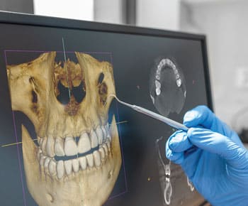
Similarly, CBCT scans can provide key information to support periodontal or implant therapy. “This technology can be used to measure the density, height and buccolingual width of the alveolar bone at any jaw location,” he explains. “It also aids evaluation of root morphology, as well as visualization of pathology, inclination of the bone, and vital anatomic structures.”
In addition, the data can be used to fabricate surgical guides that enhance the accuracy of implant placement. “Such guides can also help clinicians generate retro-engineered casts that enable the prefabrication of restorations prior to implant surgery,” Frantz says. “These restorations can be precisely engineered to improve prosthetic outcomes and delivered on the day of the operation.”
When deciding on a CBCT unit, clinicians will need to choose between regional and full-scan machines, which is often discussed in terms of field of view (FOV). This is determined by clinical use. As Frantz notes, “Large FOV images are needed by periodontists, orthodontists, oral and maxillofacial surgeons, and prosthodontists for advanced reconstruction with dental implants. However, for most other situations, a more restricted FOV is suitable — which is a more economical option that also decreases the patient’s radiation exposure.”
Another wrinkle to consider is 3D stitching, which combines two or three small FOV volumes into a larger, composite image that lets dentists examine a wider region. This can broaden the range of applications, while simultaneously enhancing flexibility, affordability and workflows. It also allows clinicians to optimize the radiation dose. “Based on current evidence, stitched composite 3D imaging appears to be accurate for diagnostic purposes,” Frantz reports.
Providers who are interested in cone beam scans should also be aware of scatter. Characterized as a key limitation to CBCT image quality, this phenomenon occurs when photons are diverted from their original path by interaction with matter. According to Frantz, scatter can manifest as streaks and other artifacts that can reduce soft tissue contrast and affect the appearance of tissue density. This possibility should always be considered when reading the scans.
Obviously, implementing CBCT imaging into daily practice involves a learning curve. Yet, where indicated, 3D radiography brings many benefits in terms of diagnosis, treatment planning and delivery — especially in complex cases, where use of this advanced technology can contribute to more predictable outcomes.
From Decisions in Dentistry. January 2023;9(1)46.
