
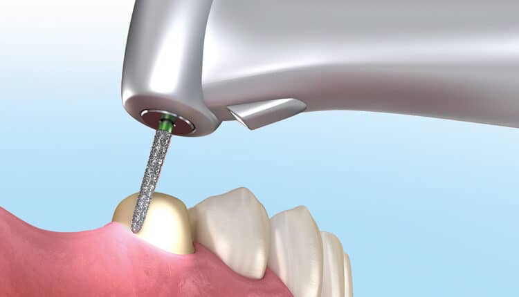
 ALEX-MIT / ISTOCK / GETTY IMAGES PLUS
Managing the Unexpected: Modification of Existing Richmond Crowns
As this case report illustrates, clinicians may encounter restorations and materials that are no longer widely used, necessitating the ability to manage the unexpected.
Oral healthcare providers often face clinical challenges, whether in the diagnostic and treatment planning stages or while completing treatment itself. Experience is always helpful in these instances, as it minimizes the chances of encountering an unfamiliar restoration or condition — especially one that warrants changes to a previously planned procedure. This case report discusses just such an instance involving the restoration of previously restored and endodontically treated teeth. The specific restorations in this case are Richmond crowns, and while this type of restoration is not used as commonly as in the past, they still exist — and must be managed when problems arise.
The restoration of endodontically treated teeth presents a restorative challenge, with a host of different techniques and materials utilized over the years. In teeth where a significant amount of coronal tooth structure is missing, some type of post is indicated in order to retain the eventual core material, which would then receive a full coverage coronal restoration. Posts were often fabricated out of metal — examples include prefabricated stainless steel or titanium, or cast gold made from a pattern formed either directly in the mouth or indirectly utilizing an impression. Posts can also be fabricated using composites reinforced with carbon or glass fibers. Core materials include amalgam, resin modified glass ionomers and composite resin, with composite resin being the most popular due to its many advantages over other materials.1 Cores can also be cast out of gold when utilizing an indirect technique in which an impression is made of the prepared root canal space and the post-core combination is subsequently cast.1
Techniques have evolved over the years, and the development of ceramics and reliable bonding systems has allowed the profession to modify the traditional approach of placing a post and core, followed by a crown. Current techniques open up the possibility of combining the post/core and crown into a single restoration known as an endo crown.2,3 Combined with chairside scanning, CAD/CAM technology lets dental teams complete all procedures in a single visit. This is an important advantage for the patient, with a significant reduction in appointment time. The ability to provide early full coverage to an endodontically treated tooth also reduces the time it is susceptible to fracture, which is always a concern when managing an endodontically treated tooth that has had significant restorative work.

One of the earliest attempts to restore an endodontically treated tooth included a single unit casting of the post, core and crown, commonly referred to as a Richmond crown. This crown was first utilized in 1878 and was described as a one-piece cast dowel and crown with a porcelain facing.4 Tylman’s5 1970 textbook describes the meticulous fabrication of a cast or soldered gold crown utilizing a dowel and porcelain facing. Tylman states these crowns were not utilized extensively, partly due to the difficulty and skill involved in fabrication. Materials and techniques improved over the years, but certain challenges remain. One technical challenge involves ensuring the internal draw required for the post fabrication, along with external draw required for the crown — all in one preparation. A disadvantage to this style of restoration lies in the difficulty encountered when a remake is necessary, whether due to material failure or caries. This was the restorative challenge presented in this case report.

CASE REPORT
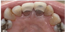
A 79-year-old female reported to the second-year clinic in the Department of Restorative Sciences at the Dental College of Georgia at Augusta University in January 2021 with the chief complaint of the unesthetic appearance of her crowns on teeth #8 and 9. She had been seen by multiple dental students since April 2003 for comprehensive care. The patient presented to the clinic in 2011 wanting to “get rid of dark circles around her crowns.” The patient did not have the finances to replace the existing crowns, so she chose to have those surfaces covered with composite. The patient had both teeth #8 and 9 treated multiple times over the next five years, with the intent of masking the underlying metal margins and improving esthetics. The results were never totally satisfactory, and the patient decided to take a more definitive approach and have the crowns replaced.

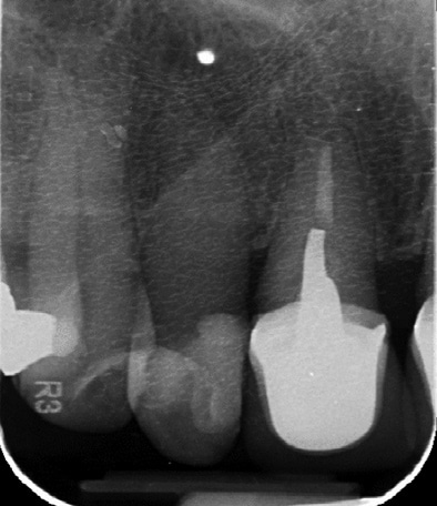
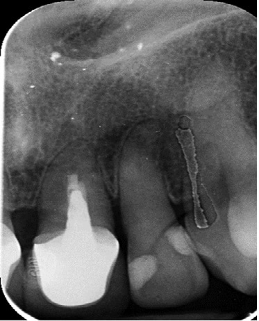
Upon reviewing the radiographs (Figure 1 and Figure 2), it was determined these teeth both had nonsurgical root canal treatment, posts and cores, and what appeared to be porcelain-fused-to-metal crowns. The patient reported this treatment initially occurred when she was 16 years old. The patient believed that she had provisional restorations on these teeth for approximately 17 years before another dentist, who did not make the original provisional restorations, realized that final restorations had not been placed. The existing crowns were estimated to be at approximately 46 years old.

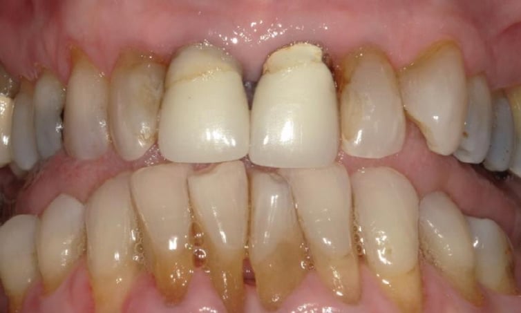
When the patient presented for definitive care for the crowns, the comprehensive exam noted periodontal disease and multiple root surface lesions that needed to be addressed prior to treating teeth #8 and 9. A number of potential treatment plans were discussed because the patient presented with multiple esthetic challenges, including rotated and malposed lateral incisors, in addition to the unesthetic crowns on teeth #8 and 9 (Figure 3, Figures 4A and 4B, and Figure 5). The patient’s chief complaint centered on the crowns, so the initial treatment plan focused on replacing those crowns.
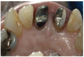
Tooth #8 had clinical and radiographic evidence of secondary caries around the distal margin. Treatment started in February 2022 on tooth #8, with the plan to remove the crowns, address the caries, and prep for new crowns. Shade C3 was chosen using the Vita Classical shade guide. As mentioned previously, the crowns were presumed to be porcelain fused to metal. Initial cuts were made on the crown in an attempt to carefully separate the crown without interfering with the existing post and core. As the preparation continued, there was no evidence of a cement line between the presumed crown, post and core evident on the radiograph. At this point, the attending faculty assisted in the preparation to ensure the initial depth cut of the bur was deep enough to reach the presumed cement line.
The distal half of the metal core fractured off in the process, and the attending faculty and student subsequently determined these were actually Richmond crowns. It was impossible to tell from clinical or radiographic evidence, but the clinical presentation at the time of treatment led to the conclusion the crowns had been fabricated as a single casting (i.e., a Richmond crown). Next, the remaining porcelain was prepared off the mesial half of the tooth so the remaining core would not fracture. The existing core was repaired by etching the surface and tooth with 35% phosphoric acid, applying a metal primer and dental adhesive to the metal core and tooth, and placing composite where necessary to complete the core.
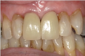
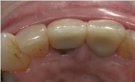
All remaining porcelain was prepped away initially using a diamond chamfer bur, and completed using a diamond shoulder bur to finalize the preparation with appropriate draw and margins for porcelain-fused-to-zirconia crowns. With the knowledge that tooth #8 was a Richmond crown, the decision was made to prepare the porcelain away from the metal on tooth #9 and create a three-quarters crown, as seen completed in Figure 6, Figure 7 and Figure 8. The preparations were completed at a subsequent appointment and final crowns were cemented a short while later (Figure 9 and Figure 10).
![Canine Richmond Crown]() Canine Richmond Crown
Canine Richmond Crown

The attending faculty author of this article had limited experience with Richmond crowns prior to the case described in this paper. The only previous experience he had in fabricating a Richmond crown involved a military working dog. The dog had experienced a fractured canine during a training exercise (Figure 13) that necessitated an endodontic procedure, followed by some type of restoration to replace the lost coronal tooth structure. Since any treatment would require sedation, the decision was made to utilize a single post-core-crown casting. After preparation of the canal space for the post, and external surface of the remaining tooth structure for the crown (Figure 14), an impression was made using polyvinylsiloxane material (Figure 15, the working cast). A casting using a nonprecious alloy was then fabricated and the crown was cemented at a subsequent appointment (Figure 16). Unfortunately, the patient fractured the crown-post during another training exercise and the tooth was restored using an amalgam core (Figure 17). In hindsight, the post probably should have been longer in order to withstand the tremendous biting force used by military dogs during training exercises.
William R. Bachand, DDS
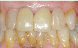
The underlying metal cores presented a challenge in masking the dark shade of the metal, but the lab assisted by applying an opaque white zirconia liquid to the intaglio of the crown that masked any underlying darkness. The unesthetic composites on teeth #7 and 10 and malalignment of 7 were then addressed with facial composite veneers using shades C2 and C3 from the Vita Classical shade guide. The shade of teeth #8 and 9 appears more yellow in the final photographs, although they seemed much closer to the shade of the lateral incisors during the try-in and cementation. The patient indicated being satisfied with the esthetics of teeth #7 to 10 (Figure 11 and Figure 12).
DISCUSSION
This patient presented with multiple challenges to the esthetics of her anterior teeth, with rotated and malposed lateral incisors, along with stained restorations and recurrent caries. The failing and unesthetic crowns on teeth #8 and 9 were her main concern, however. Multiple treatment possibilities were initially considered, including placing four crowns from teeth #7 to 10, crowns on 8 and 9, with porcelain veneers on 7 and 10, and, finally, replacing just the crowns on 8 and 9 (again, her primary concern).
According to the patient, the Richmond crowns were 46 years old. The existing endodontics and posts were considered adequate. While the endodontic treatment for tooth #9 was far from ideal, there was no evidence of periapical radiolucency and the tooth was asymptomatic. Again, according to the patient, the endodontic treatment was completed more than 60 years ago. It is unknown if the endodontic portion was retreated in 1976 when the existing crowns were completed. Endodontic retreatment would involve post removal, which could present the risk of root fracture or apical surgery. The radiographic appearance and lack of symptoms led to the decision to try to avoid involving the existing endodontic treatment and cores (if at all possible).
Although the solid post-core-crown construction was unexpected, the planned treatment was modified to address the initial caries on tooth #8, keeping the existing post and much of the core intact. The plan changed to ostensibly treat 8 as if it had a cast post and core in place, with a separate crown prepared over it.
The crown on tooth #9 had an unsightly repaired facial margin and unesthetic facial porcelain, but no caries along the margins. The decision was made to prep tooth #9 like 8, but keep the lingual margins intact since no problems were noted. The margins could have been dropped on the lingual, but the plan was to keep this preparation a bit more conservative in order to minimize any possible disturbance to the existing post and core.
When the crowns on teeth #8 and 9 were completed, the patient expressed satisfaction, but her laterals still detracted from the esthetics that could be achieved. The composite bonding on teeth #7 and 10 was then provided to complete the esthetic rehabilitation of the anterior teeth. The patient indicated she was pleased with the result.
CONCLUSION
As this case illustrates, unexpected situations can arise during care and providers must be prepared to modify existing treatment or develop alternative ways to reach a desired goal. Materials and techniques change over the years, and it would benefit practitioners to be cognizant of not only current methods of treatment, but also some that may no longer be utilized as frequently, if at all. There is a significant percentage of the U.S. population that could present with dental work that may be 30 to 50 years old. Although that says a lot about the longevity of dental care in some instances, it can also present clinical challenges.
This report describes just such a case — Richmond crowns that were not identified as such during diagnosis and treatment planning. Although the initial treatment plan did not change, the preparation designs had to be significantly modified to meet this unexpected occurrence.
The foregoing presentation also highlights some key issues regarding esthetics. The patient is always the final arbiter. Sometimes the patient can be very demanding — and dental professionals tend to be exacting, as well. In this case, the patient was pleased with the final crowns (although the shade does seem off — at least to the clinical team). She was also not as concerned about the lateral incisors, although various options to address the laterals were discussed. In the end, the patient expressed complete satisfaction with the final result and care she received.
Key Takeaways
- Clinical care can present unexpected challenges, so providers should be prepared to encounter unfamiliar restorations or conditions — especially presentations that warrant changes to previously planned treatment.
- For example, this case presentation details the retreatment of Richmond crowns, and while this type of restoration is not used as commonly as in the past, they still exist — and must be managed when problems arise.
- During the initial clinical exam, the patient’s chief concern was the unesthetic appearance of her crowns on teeth #8 and 9.
- Upon reviewing the radiographs, it was determined these teeth both had nonsurgical root canal treatment, posts and cores, and what appeared to be porcelain-fused-to-metal crowns.
- During the preparation phase, it was discovered the crowns on teeth #8 and 9 featured solid post-core-crown construction, and were, in fact, Richmond crowns.
- As this was unexpected, the planned treatment was modified to keep the existing post and much of the core intact. The plan changed to treat tooth #8 as if it had a cast post and core in place, with a separate crown prepared over it.
- The crown on tooth #9 had an unsightly repaired facial margin and unesthetic facial porcelain, but no caries along the margins. The decision was made to prep tooth #9 like 8, but keep the lingual margins intact since no problems were noted.
- As this case illustrates, unexpected situations can arise during care and providers must be prepared to modify existing treatment or develop alternative ways to reach the desired goal.
- Materials and techniques change over the years, and it would benefit practitioners to be cognizant of not only current methods of treatment, but also some that may no longer be utilized as frequently, if at all.
REFERENCES
- Hilton TJ, Ferracane JL, Broome JC. Summitt’s Fundamentals of Operative Dentistry: A Contemporary Approach. 4th ed. Hanover Park, Ill: Quintessence Publishing; 2013:562–578.
- Govare N, Contrepois M. Endocrowns: A systematic review. J Prosthet Dent. 2020;123: 411–418.
- Papalexopoulos D, Samartzi TK, Sarafianou A. A thorough analysis of the endocrown restoration: A literature review. J Contemp Dent Pract. 2021;22:422–426.
- Mishra P, Mantri SS, Deogade S, Gupta P. Richmond crown: A lost state of art. Int J Dent Health Sci. 2015;2:448–453.
- Tylman SD. Theory and Practice of Crown and Fixed Partial Prosthodontics (Bridge). 6th ed. St. Louis, Mo: CV Mosby; 1970;584–599.
From Decisions in Dentistry. December 2022;8(12)10,13-14,16.
