
Navigated Implant Placement Restores a Patient’s Smile
Through digital navigation and three-dimensional planning, a missing lower premolar was restored with pinpoint accuracy. This case highlights the power of advanced technology to deliver predictable, esthetic implant outcomes.
A woman seeking replacement of her missing lower premolar found a high-tech solution at the Center of Implant, Esthetic, and Innovative Dentistry at Indiana University School of Dentistry. Using guided navigation, the clinical team planned and executed a precise, restoratively driven implant procedure from start to finish.
A 45-year-old woman presented with a chief complaint of “I want to replace my lower missing tooth,” pointing at the lower left second premolar (#20). The medical history showed no contraindication to surgical implant placement.
An X-clip impression was taken on the lower right canine to the second premolar area. The clip was taken out and placed back into the patient’s mouth to verify that it was stable and not easily dislodged. A cone-beam computed tomography (CBCT) was then taken of the lower jaw with the clip still in the patient’s mouth. The “digital imaging and communications in medicine” (DICOM) data obtained from the CBCT were then uploaded onto the navigation system’s computer and a restoratively driven implant plan was completed.
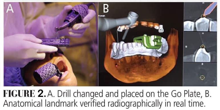
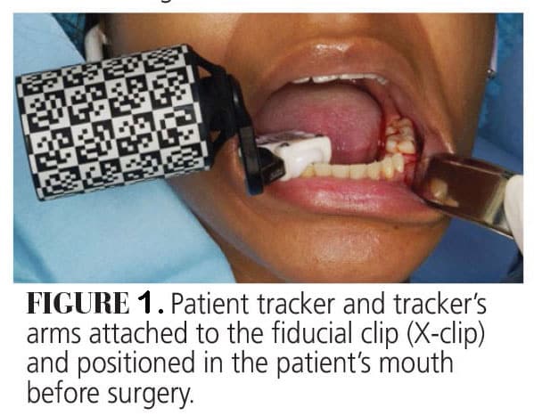
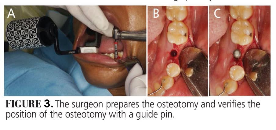
The handpiece tracker was attached to the handpiece and calibrated into the system. A green (right) tracker arm was attached to the X-clip and the patient tracker was screwed on it and a patient tracker calibration was performed. The X-clip was placed back into the patient’s mouth with the tracker attached to it (Figure 1). A full-thickness flap was reflected on the area of interest and a system check was performed prior to implant surgery and with every drill change (Figure 2). After drilling the last drill, a guide pin was placed to verify the location osteotomy (Figure 3).
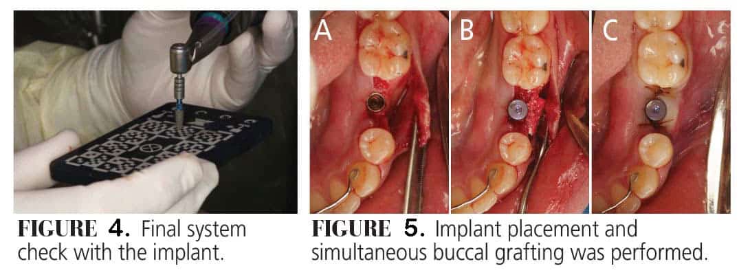
The implant was placed on the handpiece and a final system check was done (Figure 4). The implant was then placed into the prepared osteotomy site. Due to a slight buccal concavity and thin buccal bone, the surgeon opted to graft that site. A healing abutment was placed, and the site was sutured (Figure 5).
This case demonstrates how dynamic navigation systems elevate implant dentistry, offering real-time visualization, meticulous control, and the ability to adapt to intraoperative findings. The result: a stable, esthetic implant placement tailored to the patient’s unique anatomy and restorative needs.
This originally appeared in AlQallaf H, Lin SW, Polido W, Yang CC. Exploring dynamic computer-assisted implant surgery. Decisions in Dentistry. 2024;10(6):32-35.
