

Addressing the Esthetic Challenges of Implant Restorations
A patient presented with dissatisfaction over an unesthetic implant-supported crown for his maxillary central incisor, compounded by thin tissue and soft tissue deficiencies. This case highlights a comprehensive approach to soft tissue augmentation and restorative techniques to achieve a functional and esthetic result.
 Patient A sought our expertise to improve the appearance of his maxillary left central incisor, which was restored with an implant-supported crown. His concerns included the visible grayish hue of the implant due to thin tissue, chipped porcelain, and the unnatural appearance of his dentition’s shape and spacing. A detailed evaluation revealed soft tissue deficiencies, poor implant emergence profile, and tissue discrepancies that required a customized treatment plan aimed at enhancing both esthetics and function.
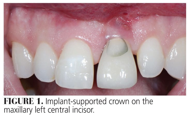
Patient A was referred to our dental office for evaluation of a single implant-supported crown that replaced his maxillary left central incisor (Figure 1). His chief complaint was the unesthetic appearance when he smiled, spaces between his teeth, and orientation and shape of his teeth. Furthermore, the patient complained about the chipped porcelain on his implant-supported crown.
Patient A’s natural tooth was lost after an endodontic procedure failed and he was treated with an immediate implant placement after tooth extraction. This entire dental treatment was performed overseas in 2007, therefore any information regarding the implant (type, size) was not available.
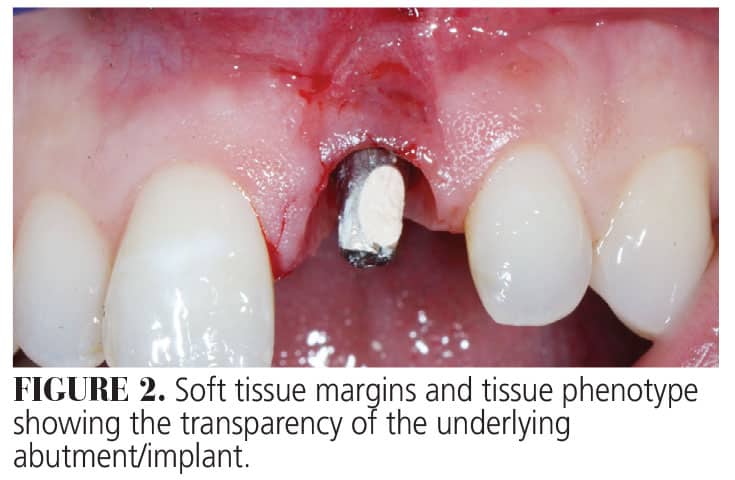
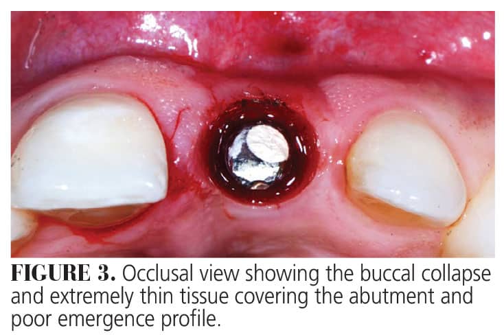
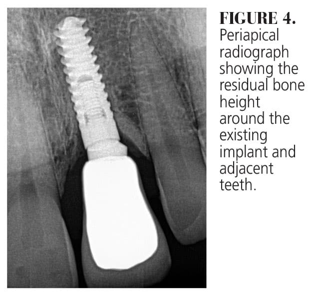
The patient’s medical history revealed no systemic contraindications for dental treatment. Clinical examination showed a thin tissue phenotype, with the gingival margin of the implant in a more apical position with respect to the adjacent natural teeth. The tissue was very thin, allowing the underlying grayish hue of the implant surface to be visible (Figure 2). In addition to the horizontal soft tissue deficiency, there was inadequate emergence profile of the implant abutment (Figure 3). Fortunately, radiographic examination determined that interproximal bone loss around the implant and the adjacent teeth was minimal. In addition, no signs of any peri-implant issues were present (Figure 4).
 The primary treatment goals were to reduce the soft tissue deficiency (vertically and horizontally), eliminate the soft tissue discrepancy between the implant restoration and the right central incisor, and place a new crown over the implant. The plan also included changing position and angulation of the maxillary anterior teeth as well as their shape as the patient desired squarer-looking teeth. Furthermore, the tissue thickness over the implant was to be increased to mask the implant/abutment.
This case underscores the importance of addressing both biological and esthetic aspects of implant dentistry to meet patient expectations. Through careful planning and a multidisciplinary approach, we achieved a harmonious, natural-looking result, restoring the patient’s confidence in his smile.
This information originally appeared in Boeriu S, Hottel TL, Chirla C, Chirla P. Recession defects on a maxillary single tooth implant. Decisions in Dentistry. March 2024;10(2):16-19.
