

CBCT Transforms Implant Planning and Periodontal Care
Cone beam computed tomography (CBCT) delivers unparalleled imaging for dental implant planning and periodontal care. Discover how this cutting-edge technology balances diagnostic value with radiation safety to set new standards in dental practice.
Cone beam computed tomography (CBCT) was introduced to the dental community more than 20 years ago.1 This technology gathers quality images of the dentition and surrounding structures that provide valuable, real-time information for dental implant planning. More recently, CBCT imaging has been found to aid in the diagnosis and treatment of various periodontal conditions — and these scans are captured more quickly and easily than conventional medical computed tomography.2 But this added radiation dose, and how it compares with conventional diagnostic imaging, should be considered.
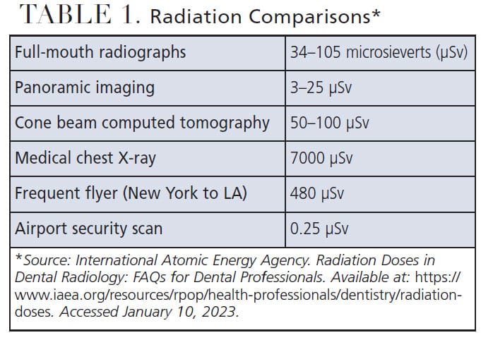
Table 1 shows comparative dosage levels from various radiation sources. It is important to remember that no matter how low the radiation dose, it is deemed excessive if it does not improve therapeutic outcomes.3,4 It is hoped this review will provide context for the possible application of cone beam imaging in dental implant planning and placement, as well as when diagnosing and treating periodontal diseases.
Professional dental associations have authored opinion or position papers on the role of CBCT in dental implant surgical planning. The American Academy of Oral and Maxillofacial Radiology suggests the preoperative diagnostic phase of implant planning should use cross-sectional imaging, as it will provide diagnostic value at a tolerable radiation dose.5 In some countries, acquiring a CBCT for implant planning purposes is already the standard of care.6 In 2017, the American Academy of Periodontology investigated whether cone beam imaging should replace standard two-dimensional films for patients requiring dental implant surgery.7
References
- Eke PI, Thornton‐Evans GO, Dye BA, Genco RJ. Advances in surveillance of periodontitis: The Centers for Disease Control and Prevention periodontal disease surveillance project. J Periodontol. 2012;83:1337–1342.
- Acar B, Kamburoglu K. Use of cone beam computed tomography in periodontology. World J Radiol. 2014;6:139–147.
- American Dental Association Council on Scientific Affairs. The use of cone-beam computed tomography in dentistry: an advisory statement from the American Dental Association Council on Scientific Affairs. J Am Dent Assoc. 2012;143:899–902.
- Walter C, Weiger R, Zitmann N. Accuracy of three-dimensional imaging in assessing maxillary molar furcation involvement. J Clin Periodontol. 2010;37:436–441.
- Tyndall DA, Price JB, Tetradis S, Ganz SD. Position statement of the American Academy of Oral and Maxillofacial Radiology on selection criteria for the use of radiology in dental implantology with emphasis on cone beam computed tomography. Oral Surg Oral Med Oral Pathol Oral Radiol. 2012;113:817–826.
- Mandelaris GA, Scheyer ET, Evans M, et al. American Academy of Periodontology best evidence consensus statement on selected oral applications for cone-beam computed tomography. J Periodontol. 2017;88:939–945.
- McAllister B, Eshraghi VT. Commentary: cone-beam computed tomography — an essential technology for management of complex periodontics and implant cases. J Periodontol. 2017;88:937–938.
