

A Case Report on Effective Treatment for a Mucogingival Defect
An older adult with severe interdental attachment loss and gingival recession underwent a procedure to increase the zone of attached gingiva. The treatment not only improved vestibular depth and soft tissue phenotype but also alleviated inflammation and sensitivity, supporting better oral hygiene practices.
In cases of severe periodontal attachment loss, addressing gingival health can be particularly challenging. This case report explores the surgical management of an older adult with significant gingival recession, inflammation, and sensitivity due to a lack of attached gingiva. The primary goal was to enhance the zone of attached gingiva and prevent further recession while promoting better oral hygiene.
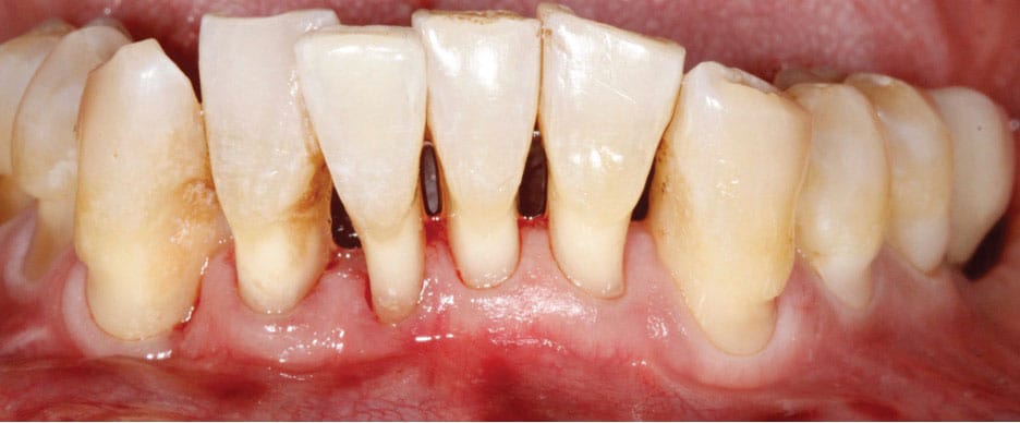
Figure 1A depicts an older adult patient with a history of periodontitis resulting in generalized interdental attachment loss, gingival recession, and a lack of attached gingiva that resulted in a shallow vestibule. The patient’s lack of attached gingiva led to persistent inflammation and sensitivity around the gingiva. Due to the extent of interdental attachment loss, the predictability of obtaining root coverage was significantly reduced. Therefore, the primary goal of the procedure was to increase the zone of attached gingiva, thereby improving vestibular depth and preventing her recession from progressing. This would also support the patient’s oral hygiene efforts.
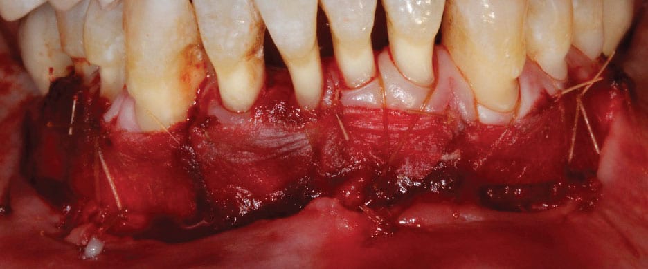
The recipient site was prepared by performing a split-thickness dissection from teeth #19 to 30. The alveolar mucosa was sutured to the periosteum using resorbable chromic gut sutures. Next, xenogeneic collagen matrix was secured to the recipient site with resorbable 5-0 chromic gut sutures (Figure 1B). A surgical dressing was placed over the recipient site because the graft was secured to the recipient bed and left exposed. This also protected the area from external irritation or trauma, allowing the site to heal uneventfully.
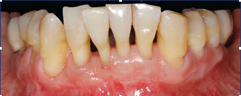
Post-operative instructions included a soft diet, no brushing and flossing in the area, and utilization of an antibacterial mouthrinse. The surgical dressing and remaining sutures were removed after two weeks. At the 6-month follow-up appointment, a wide zone of attached gingiva was observed extending from teeth #19 to 30. Restoration of vestibular depth and improvement in the patient’s soft tissue phenotype were also noted (Figure 1C). Gingival sensitivity and tissue susceptibility to inflammation were significantly improved following the procedure.
This case highlights the success of a targeted surgical approach in addressing gingival recession and inflammation in a periodontally compromised patient. The results demonstrate the importance of increasing attached gingiva to improve oral health outcomes, enhance patient comfort, and support long-term stability.
This information originally appeared in Shaikh S, Regahi P. Soft tissue graft alternatives for treating mucogingival defects. Decisions in Dentistry. 2023;9(1):26-29.
