
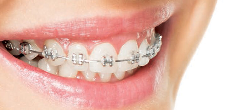
Insights Into Surgically Assisted Osteogenic Orthodontics
This surgical procedure will help accelerate tooth movement and significantly decrease orthodontic treatment times.
This surgical procedure will help accelerate tooth movement and significantly decrease orthodontic treatment times
In the late 1990s, as increasing numbers of adults sought orthodontic therapy, dentists were faced with two major problems. These patients want to spend a minimal amount of time in braces, and therapists can no longer use facial growth to their advantage.
In response, two brothers — one an orthodontist and the other a periodontist — fused well-known surgical techniques into a single therapy to help address these concerns. The Wilcko brothers called their procedure accelerated osteogenic orthodontics (AOO). Also known as Wilckodontics,1 this is a surgical procedure designed to enhance orthodontic outcomes. The concept involves scoring of the alveolar bone (i.e., a corticotomy), accompanied by hard tissue grafting and, when appropriate, soft tissue grafting. Their original goal was to decrease the time needed for tooth movement using fixed orthodontic appliances. They also reported that one of the significant advantages was a reduction in apical resorption following therapy. This finding has been reported elsewhere.2
Fixed orthodontic appliances are placed before surgical procedures are performed. The day of the surgery, the orthodontic wire is removed. Full thickness mucoperiosteal flaps are raised in the areas to be treated. The Wilcko’s original approach called for elevation of facial and lingual (palatal) flaps. Corticotomies were performed between and apical to the teeth to be moved on the facial, and between the roots of the teeth on the palatal (lingual). Palatal cuts were not beyond the apices.
Bone allografts (decalcified freeze-dried and bone allograft) and bovine bone (xenografts) were mixed and wetted with clindamycin placed over the surgical sites. The flaps were then replaced and sutured. Tooth movement is initiated by replacing the orthodontic wire after surgery (normally within 48 hours). Tooth movement has been demonstrated to occur histologically after three days.3 The technique was later modified by the Wilckos to include different types of allografts, and xenografts along with the thinning of dense facial or lingual (palatal) bone.
TERMINOLOGY DISTINCTIONS
This technique relies on the bone’s response to surgical trauma. It is important to define and recognize the differences in the terminology used in the literature to describe the way in which the bone is treated to obtain accelerated tooth movement. A corticotomy has been defined as a surgical procedure in which only the cortical bone is cut or mechanically altered, while an osteotomy is a surgical cut through both the cortical and medullary bone.4 Wang and coworkers demonstrated in the rat that corticotomies produced transient bone resorption around dental roots, which was not observed with osteotomies.5 Wounding of the bone, as described above, results in simultaneous stimulation of anabolic and catabolic activities, and produces what has been called the regional accelerated phenomenon (RAP).6 This is clinically relevant because RAP results in accelerated bone turnover and a decrease in regional bone densities in the areas of surgical intervention. Following the corticotomy, there is an increase in osteoclastic activity, and increased number of osteoclasts, less trabecular bone surface (osteopenia), as well as a wide periodontal ligament space around the affected teeth.7 It is accepted that bone grafting both stabilizes the dentition, and also acts as a scaffold to reduce the probability of gingival recession. In one study, computerized tomography was used to measure pre- and postcorticotomy thickness of the facial alveolar bone in 20 patients. The study’s conclusion was that, in all cases, corticotomies increased the facial alveolar width.8 In addition to increased apposition of bone seen in the surgical site, the hypothesis was that RAP allows the teeth to move faster and further than traditional nonsurgical orthodontic therapy.8
The question of whether or not corticotomies increased the amount of tooth movement has been studied by multiple groups. Studying dogs, Sanjideh and colleagues found increased mandibular tooth movement for a corticotomies group (versus a control group) between 22 and 25 days. They also found that mesial-distal movement of teeth in the mandible was increased approximately two times by using corticotomies.9 Current opinion is that the rapid movement produced diminishes after the first two to three months in dogs.9 It has been suggested that the window of opportunity is four to six months for humans. Similar studies by other groups found similar results. One group studied the effect of a second corticotomy on tooth movement and found that there was little additional benefit from the second surgery.10 Most of the accelerated movement occurs within 60 to 90 days following surgery.11
An examination of treatment time, studied in dogs, found that mesial distal movement was four times faster in the maxilla and two times faster in the mandible when corticotomies were used compared with controls.10 Similar results have been shown in humans. A recent systematic review suggests that initial movement is two to three times faster in humans, with deceleration of movement seen between four to six months.12
Some of the early goals of this therapy have since been modified, as the emphasis has shifted from rapidity of tooth movement to increasing the distance a tooth can be moved and still retain a stable, increased alveolar housing. Amit et al proposed a modification to the technique in which the corticotomies do not penetrate the trabecular bone. The technique proposed was termed periodontal accelerated osteogenic orthodontics (PAOO).13 The PAOO authors recommend decortication, which refers to the removal of the cortical portion of the alveolar bone on the buccal and lingual/palatal plates. Additionally, bone grafting is used to provide an increased net alveolar volume after orthodontic treatment.
SURGICALLY ASSISTED OSTEOGENIC ORTHODONTICS
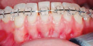
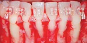
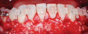
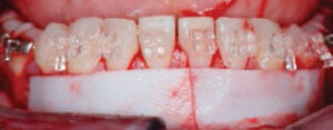
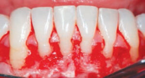
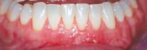
In this article, we will discuss a different approach that involves full thickness flaps and corticotomies — but now only on the facial aspect of the teeth to be moved. Corticotomies are performed using thin piezoelectric saws under copious irrigation. This minimizes the trauma to the tissues and facilitates the corticotomies in narrow, interradicular bone spaces. The corticotomies penetrate the facial cortical bone on the mesial and distal aspects, as well as 5 mm apical to the apices of the teeth to be moved. All these calculations are made using cone beam computed tomography (CBCT). Three-dimensional CBCT imaging allows precise estimations of presurgical bone volume and teeth positioning, which helps prevent injury to the roots or other adjacent vital structures, such as nerve canals or the maxillary sinuses.14 Bone allograft is placed directly over the corticotomies. Allografts contain bone morphogenetic proteins, which recruit pre-osteoblast cells in the wound and induce new bone formation. The bone graft is overlaid by a deproteinized bone xenograft, which is osteopromotive and has a lower rate of resorption than bone allografts. This xenograft helps to enhance and maintain the alveolar bone volume for a longer period because its resorption rate is slower than the allograft’s. Both grafting materials are mixed with enamel matrix derivatives (EMDs) before placement, as EMDs have known biomimetic properties that enhance cell proliferation, as well as hard and soft tissue healing. The grafted area is then covered with a resorbable collagen membrane. In areas of deficient attached/keratinized tissue, subepithelial connective tissue grafts can be overlaid at the deficient areas during the same surgical procedure. These modifications increase our ability to move teeth and create a more stable environment. This article will use the term “surgically assisted osteogenic orthodontics” (SAOO) because we believe that increased speed of tooth movement is helpful, but secondary to the enhancement of the alveolar housing seen with this technique (Figure 1 through Figure 6B). By changing the periodontium biotype/phenotype, esthetics are improved and a significant change in profile is seen in most patients. The bone augmentation has been seen to be stable, as evidenced by CBCTs obtained to two years after the procedure.
long-lasting collagen resorbable membrane.
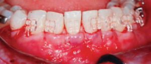
WOUND STABILITY AND BLOOD SUPPLY
An important fact about SAOO is that by maintaining an intact blood supply on the lingual/palatal, clinicians can obtain greater wound stability. Blood supply and wound stability are critical for bone healing. Because there is no lingual flap elevation, the periosteum is not severed and periosteal regenerative properties are preserved. For this reason, the initial incision is done at the base of the papillae on the buccal, maintaining the integrity of the papillae. By taking into consideration important bone healing principles, SAOO increases the amount of tooth movement that can be obtained with the orthodontic therapy. Therefore, it widens the spectra of malocclusion cases that can be treated without the need of more aggressive surgical procedures. It has been reported that in three planes of space, movement of the teeth can be increased from 2 mm to 3 mm using traditional nonsurgical orthodontic therapy, versus 4 mm to 6 mm using corticotomies. Many cases that would originally have been treated using orthognathic surgery (usually a hospital procedure) can now be done in-office. The RAP induced by the SAAO results in faster movement, decreasing treatment times by more than half in most cases. This reduces morbidity, treatment duration and expense for the patient.
key takeaways
- Accelerated osteogenic orthodontics is a surgical procedure that involves scoring of the alveolar bone (i.e., a corticotomy), accompanied by hard tissue grafting and, when appropriate, soft tissue grafting.
- This therapy decreases the time needed for tooth movement using fixed orthodontic appliances.
- Some of the early goals have since been modified, as the emphasis has shifted from rapidity of tooth movement to increasing the distance a tooth can be moved and still retain a stable, increased alveolar housing.
- By changing the periodontium biotype/phenotype, a modified procedure — known as surgically assisted osteogenic orthodontics — improves esthetics and, in most patients, results in a significant change in profile.
CONTRAINDICATIONS
Contraindications to these procedures include the usual list of systemic reasons not to do surgery. Specific contraindications include long-term use of corticosteroids, bone resorption inhibitors with known risk of osteonecrosis of the jaw, history of head and neck radiation, and active infections of endodontic and periodontal origin.
The early healing following these procedures includes edema, which usually persists for two to three days, bleeding (in trace amounts) in the saliva for 24 to 48 hours, and discomfort. This discomfort is normally treated using a combination of ibuprofen and acetaminophen every six to eight hours. The protocol also includes the use of systemic antibiotics starting the day before the procedure, followed by an eight-day course. Less frequently seen risks include damage to the involved teeth, altered healing and nerve damage.
General indications for SAOO include a patient’s desire for enhanced facial esthetics, a thin periodontium (both soft and hard tissue), the need for increased tooth movement compared to nonsurgical approaches, areas of gingival recession around the teeth to be moved, and the desire to avoid orthognathic surgery and decrease the need for extractions.
Specific clinical indications include cross bites, crowded anterior teeth, extruded teeth, narrow buccal corridors, the desire for faster tooth movement, and the desire to do the surgery as an office procedure. The treatment planning phase should include collaboration between the orthodontist, periodontist and patient. Precise goals and limitations should be established and discussed during the presurgical meetings. Once the orthodontic plan is defined, the fixed appliances are bonded and teeth are initially aligned. It is important to notice this is a staged approach that differs from the PAOO technique. The day of the surgery, the arch wires are removed to facilitate the surgical procedure. The patient is seen by the orthodontist soon after surgery to activate the tooth movement. Orthodontic movement is activated weekly during the first two months to take advantage of RAP. Increased mobility is a desirable outcome and should be expected. Teeth tend to be more stable after the third month following SAOO.
At present, there is no human histologic data on SAOO. Animal histology, radiographic evidence and clinical experience, however, indicate that RAP does occur and routinely results in an increase in the facial-lingual dimension of the alveolar housing. It has been hypothesized that this will stabilize teeth and reduce the probability of gingival recession in the future.
CLINICAL DISCUSSION
The surgical procedures described above have changed the profession’s approaches to select orthodontic cases. Other procedures have been used to accelerate dental movement, including vitamin D therapies, vibratory forces, pulsed electromagnetic fields, electrical current and low laser radiation. Some increase in movement rates has been observed with these procedures.15 Other approaches published in the literature include the use of local injection of biomodulators and gene therapy. Basic research in these areas is promising, but still not available.16 In general, RAP has been used to potentiate hard tissue remodeling by several proponents of surgery in conjunction with orthodontics. Some of these techniques include corticotomies, piezocision, periodontal distraction, mocortical tooth dislocation and ligament distraction, alveocentesis (micro-osteoperforations) and corticision. All these proposed techniques have a low-to-moderate level of evidence because they are based on case reports without control groups.15 Some corticotomy-assisted orthodontic procedures have low acceptance from patients. Fear of surgery was the main reason for not accepting the surgical procedures.17
The SAOO procedure allows tooth movement into areas of minimal or no bone, and produces clinical and radiographic signs of a more robust bony housing in areas of once deficient bone. Histological studies in animals have demonstrated that corticotomies, followed by grafting, induced large amounts of new bone formation.18 Also, histological observation in canines has suggested this procedure can stimulate periodontal tissue reestablishment and the formation of a mesenchymal matrix, facilitating an even bone surface.18 This approach has been found to be helpful in deep cross bites, narrow buccal corridors, and for the intrusion of teeth. Additional benefit has been found for the occasional tooth that resists mechanical movement using normal orthodontic forces. Instead of applying more and more force, often with no positive benefit and the possibility of increased resorption, corticotomies routinely allow movement of these teeth (in most cases). In our patient population, the acceptance rate is high, which may be related to involving the patient in the treatment planning phase. Patient education is an important component to this therapy because it contributes to patient compliance and therapeutic success. The disadvantages of this procedure are additional surgical costs, pain and swelling, and the time required for planning and follow-up after surgery. The main advantages, besides additional bone support, include reduced treatment time, low relapse rates, less need for extraoral appliances, and reduced issues with root resorption.
Less invasive alternatives seem to be well accepted by the orthodontics community. Some of these include the use of flapless corticotomies or minimally invasive surgery. Piezocision, for example, is a flapless procedure combining piezo surgical cortical micro-incisions. Proponents claim this technique can locally affect alveolar bone metabolism in order to obtain rapid orthodontic results. This minimally invasive approach may be appropriate in cases in which little or no bone augmentation is necessary.19
Another published technique is the so called monocortical tooth dislocation and ligament distraction. Proponents propose the use of a microsurgical corticotomy around each tooth, buccal monocortical tooth dislocation, and palatal ligament distraction movement, followed by the application of mechanical forces.20
Micro-osteoperforation (also called alveocentesis) has been used for the treatment of rotation, molar uprighting, presurgical orthodontics, and in cases of intrusion and crowding with some success.21,22
Even though corticotomy-based techniques are predicated on the sophisticated mechanisms of bone modeling and remodeling, the level of evidence is low, as the majority of publications are case reports with small number of subjects and lacking control groups.23
CONCLUSION
Evidence gathered over the last decade suggests that a unique surgical approach to orthodontic movement can enhance outcomes, as well as reduce treatment time. While further data is needed, the results to date using this approach in select cases has been shown to enhance short- and long-term orthodontic outcomes.
References
- Wilcko WM, Wilcko MT, Bouquot JE, Ferguson DJ. Rapid orthodontics with alveolar reshaping: two case reports of decrowding. Int J Periodont Res. 2001;21(1):9–19.
- Hassan AH, Al-Saeed SH, Al-Maghlouth BA, Bahammam MA, Linjawi AI, El-Bilay TH. Corticotomy-assisted orthodontis treatment: a systemic review of the biological basis and clinical effectiveness. Saudi Med J. 2015;36(7):794–801.
- Choi HJ, Lee DY, Kim TW. Dynamics of alloplastic bone grafts on an early stage of corticotomy-facilitated orthodontic tooth movement in beagle dogs. BioMed Research International. 2014;doi:10.1155/2014/417541.
- Murphy KG, Wilcko TM, Wilcko WM, and Ferguson DJ. Periodontal accelerated osteogenic orthodontics: a description of the surgical technique. J Oral Maxillofac Surg. 2009;67(10):2160–2166.
- Lei Wang L, Lee W, Lei DL, Liu YP, Yamashita DD, Yen SL. Tisssue responses in corticotomy- and osteotomy-assisted tooth movements in rats: Histology and immunostaining. Am J Orthod Dentofacial Orthop. 2009;136:770.e1–e11.
- Frost H M. The regional accelerated phenomenon. Orthop Clin N Am. 1981;12:725–726.
- Sebaoun JD, Kantarci A, Turner JW, Carvalho, Roberto S, Van Dyke, et al. Modeling of trabecular bone and lamina dura following selective alveolar decortication in rats. J Periodontol. 2008;79(9):1679–1688.
- Bhattacharya P, et al. Assessment of corticotomy facilitated tooth movement and changes in alveolar bone thickness — a CT scan study. J. Clin Diag Res. 2014;8(10) ZC26–ZC30.
- Sanjideh PA, Rossouw PE, Campbell PM, Opperman LA, Buschang PH. Tooth movements in foxhounds after one or two alveolar corticotomies. Eur J Orthodont. 2009;32(1):106–113.
- Iino S, Sakoda S, Ito G, Nishimori T, Ikeda T, Miyawaki S. Acceleration of orthodontic tooth movement by alveolar corticotomy in the dog. Am J Orthod Dentofacial Orthop. 2007;131(4):448.
- Wilcko MT, Wilcko WM, Pulver JJ, Bissada NF, Bouquot JE. Accelerated osteogenic orthodontics technique: a 1-stage surgically facilitated rapid orthodontic technique with alveolar augmentation. J Oral Maxillofac Surg. 2009;67(10):2149–2159.
- Kalemaj Z, DebernardI CL, Buti J. Efficacy of surgical and non-surgical interventions on accelerating orthodontic tooth movement: A systematic review Eur J Oral Implantol. 2015;8(1):9–24.
- Amit G, JPS K, Pankaj B, Suchinder S, Parul B. Periodontally accelerated osteogenic orthodontics (PAOO) — a review. J Clin Exp Dent. 2012;4(5):e292–e296.
- Sahai S. Recent advances in imaging technologies in implant dentistry. J Int Clin Dent Res Organ. 2015;7(1):S19–S26.
- Camacho AD, Velasquez Cujar SA. Dental movement acceleration: literature review by an alternative scientific evidence method. World J Methodol. 2014;4(3):151–162.
- Andrade I Jr, Sousa AB, da Silva GG. New therapeutic modalities to modulate orthodontic tooth movement. Dental Press J Orthod. 2014;19(6)123–133.
- Zawawi KH. Patients’ acceptance of cortocotomy-assisted orthodontics. Patient Prefer Adherence. 2015;9:1153–1158.
- Lee DY, Ahn HW, Herr Y, Kwon YH, Kim SH, Kim EC. Periodontal responses to augmented corticotomy with collagen membrane application during orthodontic buccal tipping in dogs. Biomed Res Int. 2014;doi.org/10.1155/2014/873918.
- Dibart S, Sebaoun JD, Surmenian J. Piezocision: a minimally invasive, periodontally accelerated orthodontic tooth movement procedure. Compend Contin Educ Dent. 2009; 30(6):342–344,346,348–350.
- Vercellotti T, Podesta A. Orthodontic microsurgery: a new surgically guided technique for dental movement. Int J Periodontics Restorative Dent. 2007;27(4):325–331.
- Nicozisis J. Accelerated orthodontics with alveocentesis. Princeton Orthodontics. Clin Orthod. 2012;19:1–4.
- Teixeira CC, Khoo E, Tran J, Chartres I, Liu Y, Thant LM, Khabensky I, Gart LP, Cisneros G, Alikhani M. Cytokine expression and accelerated tooth movement. J Dent Res. 2010;89(10):1135–1141.
- Camacho AD, Velásquez Cujar SAV. Dental movement acceleration: Literature review by an alternative scientific evidence method. World J Methodol. 2014;4(3):151–162.
![The controversy around adding fluoride to drinking water was recently highlighted by an article in the Wall Street Journal, which addressed its possible negative neurological effects on children.[1] The article cited recent studies on what levels of fluoride are needed to protect teeth without risking possible cognitive harm to prenatal children and infants. Link in bio.
[1] Data on link.
---
#dentistry #dentist #dental #smile #dentista #teeth #cosmeticdentistry #dentistryworld #dentalphotography #odonto #tooth #dentistrylife #orthodontics #dentalcare #dentalhygienist #dentalimplants #oralhealth #veneers #dentalstudent #dentalassistant #dentalclinic #dentistlife #dentalhygiene #teethwhitening #oralsurgery #dds #endodontics #continuingeducation #education](http://decisionsindentistry.com/wp-content/plugins/instagram-feed/img/placeholder.png)