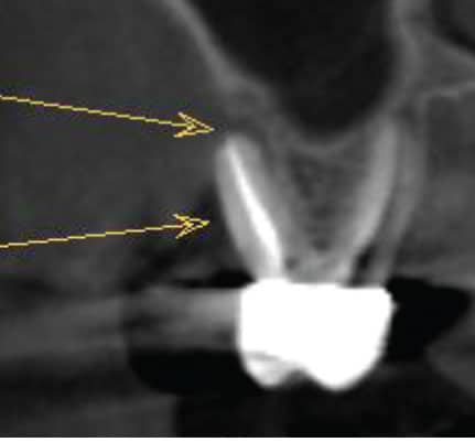
Return to "Utility of Cone Beam Imaging in Periodontics And Implant Therapy" FIGURE 2. Cone beam computed tomography showing tooth #3 and advanced loss of buccal plate. This bone destruction was not appreciated on conventional periapical radiographs, and the periodontal probing was within normal limits. Next Previous