

Seeing Beyond the Surface With Cone-Beam CT
From implant planning to airway analysis, cone-beam computed tomography is transforming dental diagnostics by revealing anatomical details that traditional radiographs can miss.
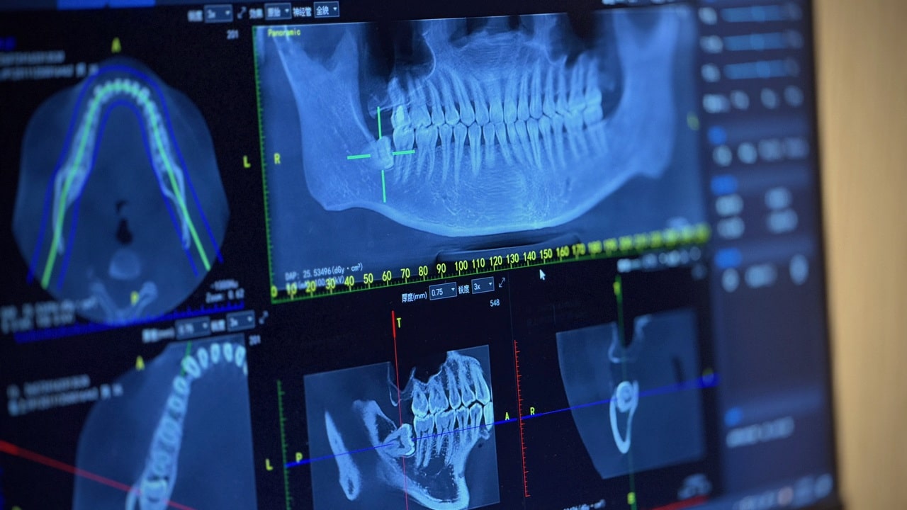
What is the primary advantage of cone-beam computed tomography (CBCT) over conventional two-dimensional (2D) dental radiography?
 Shawn Tong / iStock / Getty Images Plus
Correct!
Wrong!
CBCT provides 3D images that allow clinicians to visualize complex anatomical structures and pathologies that may be obscured by superimposition in traditional 2D radiographs.
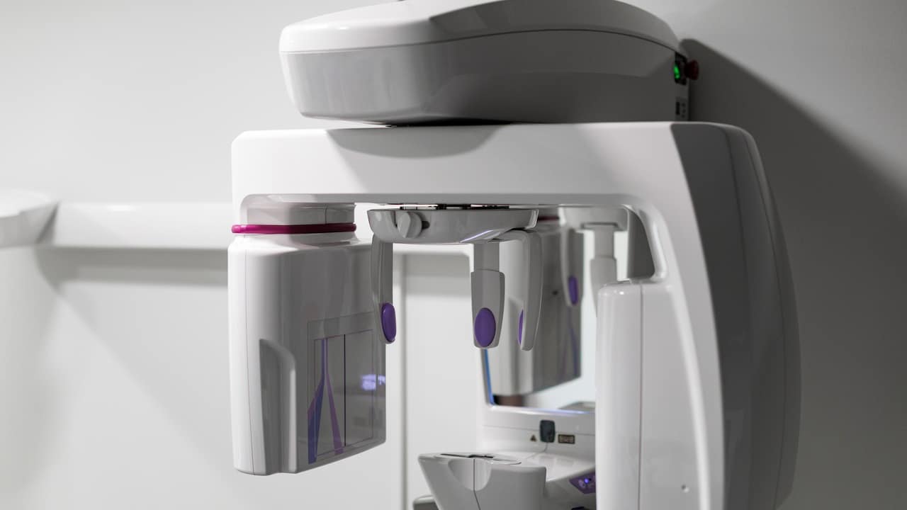
Which principle guides clinicians in selecting the appropriate field of view and resolution when obtaining CBCT images?
 ugis bralens / iStock / Getty Images Plus
Correct!
Wrong!
ALADA emphasizes selecting a field of view and image resolution that are as low as diagnostically acceptable to minimize radiation exposure while still obtaining the information needed for diagnosis.
Which imaging modality is preferred for evaluating the soft-tissue components of the temporomandibular joint (TMJ)?
 Silver Place / iStock / Getty Images Plus
Correct!
Wrong!
Although CBCT provides excellent visualization of the osseous components of the TMJ, its poor soft-tissue contrast limits its ability to evaluate soft-tissue structures. MRI is the preferred modality for assessing these components.
Seeing Beyond the Surface With Cone-Beam CT
Congratulations, you scored 100%! You really know your stuff.