
Do Probe Markings Impact Implant Health?
A recent study investigates whether probe color markings degrade through sterilization, wear off during implant probing, and transfer onto implant surfaces. Findings suggest that even gentle probing can cause color transfer, raising concerns about its potential impact on implant health and biocompatibility.
To evaluate the degradation of probe markings and the impact of color loss on implant health, a study was conducted to address the following questions:
- Do color markers deteriorate due to cleaning and sterilization?
- Does probing the implant or abutment cause the colored paint to wear off?
- How much force is needed to transfer colored material onto various implant and abutment surfaces, and how does this compare to the clinically recommended peri-implant probing force?
Click here to read the previous Perio Update that detailed the materials and methods as well as initial results.
Both probes appeared similar, however, probe A had a smoother surface and the green marker color appeared darker. This may be due to thicker paint or different paint material (Figures 1and 2). According to the manufacturer’s information, the proprietary colors are food colorants.1 The colored probes exhibited excellent resistance to wiping with isopropanol and steam autoclaving, indicating their durability and robust nature. The resilience to dissolution both in alcohol and steam autoclaving suggests that the colorant may remain in situ if retained on any implant materials.
The 2017 World Workshop on the Classification of Periodontal and Peri-Implant Diseases and Conditions defined implant health as having an absence of visible signs of inflammation and bleeding on probing. Peri-implant mucositis, characterized by bleeding on probing and visual inflammation, is considered a precursor to peri-implantitis. However, the use of terms like “gentle probing” in the classification raises concerns, as it lacks quantifiability.2

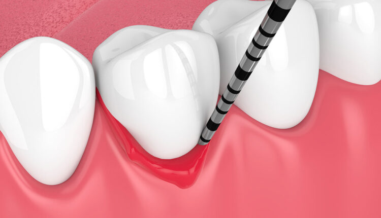
Cha et al3 conducted a study to determine the appropriate force for probing around dental implants, finding a desired pressure of 119 Ncm2. This pressure equates to a plastic probe with a 0.6 mm tip and 0.34 N of force, or 3.4 times more force than that used in this study. During initial tests, it was observed that the green color was removed due to wear at the minimal force of 0.1 N. All the tests showed that the green pigment was being worn away, which was clearly visualized on the implants and most of the abutments. For the titanium-based abutments, which had a smooth surface, SEM images were necessary to detect minute deposits and the exposure of the white probe core material after multiple passes.
The tested implant surfaces all had rough textures designed to enhance integration. This is particularly relevant as remnants of color are likely to be retained even when probed with “gentle force” of around 0.1 N. It was apparent that the test materials were harder than the probe core material, as seen from the wear on the tip of the probe after the color had been removed. However, the degree of wear was not quantified. The absence of fluid lubrication may also have affected wear and debris transfer.
The study used a force of approximately 0.1 N directed toward the probe’s tip at 90°. Although this scenario might be uncommon in clinical situations, a calculation was performed to determine the glancing angle the probe could make to mimic a 0.1 N force. Any angle greater than 17° between the implant and probe would result in color transfer when using the recommended probing force of 0.34 N on the plastic probe.3
Further investigation is required to understand the composition of the debris and its potential impact on biocompatibility with host epithelial cell attachment. Contamination of abutment surfaces has been shown to compromise the epithelial attachment of the peri-implant mucosa and inhibit the adhesion of gingival fibroblasts, ultimately leading to apical migration of the junctional epithelium.4
The study had some limitations, including the design of the probing model, use in a dry field, lack of a host peri-implant environment, material limitations, and the challenge of mimicking human factors, such as controlling and maintaining probing pressures. The true minimum threshold of debris could not be measured due to the model used which was set at a minimum force of 0.1 N due to the weight of the holding apparatus. The hardness values and surface roughness were not measured for the materials used. Further investigation into the potential effects of these implant-retained remnants should be undertaken.
Within the limitations of this study, color markings on the plastic probes were not being degraded by the action of chemical cleaning and sterilization. At a force (0.1 N) deemed considerably lower than recommended for clinical probing, colored paint contamination occurred due to frictional wear on the implants and abutments. Further investigation is required to understand the composition of the paint and plastic debris and its potential interaction and biocompatibility with host epithelial cell attachment.
References
- Fakhravar B, Khocht A, Jeffries SR, Suzuki JB. Probing and scaling instrumentation on implant abutment surfaces: an in vitro study. Implant Dent. 2012;21:311-316.
- Berglundh T, Armitage G, Araujo MG, Avila-Ortiz G, Blanco J. Peri-implant diseases and conditions: Consensus report of workgroup 4 of the 2017 World Workshop on the Classification of Periodontal and Peri-Implant Diseases and Conditions. J Periodontol. 2018;89:S313-S318.
- Cha J, Wadhwani C, Wang M, Hokett SD, Katancik J. Instrument selection and application used to probe dental implants. Int J Oral Maxillofac Implants. 2019;34:115-123.
- Guo T, Gulati K, Arora H, Han P, Fournier B, Ivanovski S. Race to invade: Understanding soft tissue integration at the transmucosal region of titanium dental implants. Dent Mater. 2021;37:816-831.
This information originally appeared in Kim SJ, Wadhwani C, Ferracane J, O’Brien R, Katancik JA. The impact of dental probe wear on implant health. Decisions in Dentistry. 2024;10(4):32-35.
