
Digital Dentistry Begins With Intraoral Scanning
Adopting intraoral scanning technology increases treatment efficiency and helps improve clinical outcomes.
The adoption of digital technology in the dental field is changing many clinical protocols, making cumbersome processes easy and the impossible possible. I believe these are positive changes for easier and more convenient clinical workflows. Digital dentistry begins with the digitization of information needed for treatment, and it is important to convert such data into digital form as accurately as possible. An intraoral scanner can be deemed the most suitable input device for digital workflow, as it directly and immediately digitizes a patient’s intraoral information, including teeth, soft tissue, and the occlusal relationship.
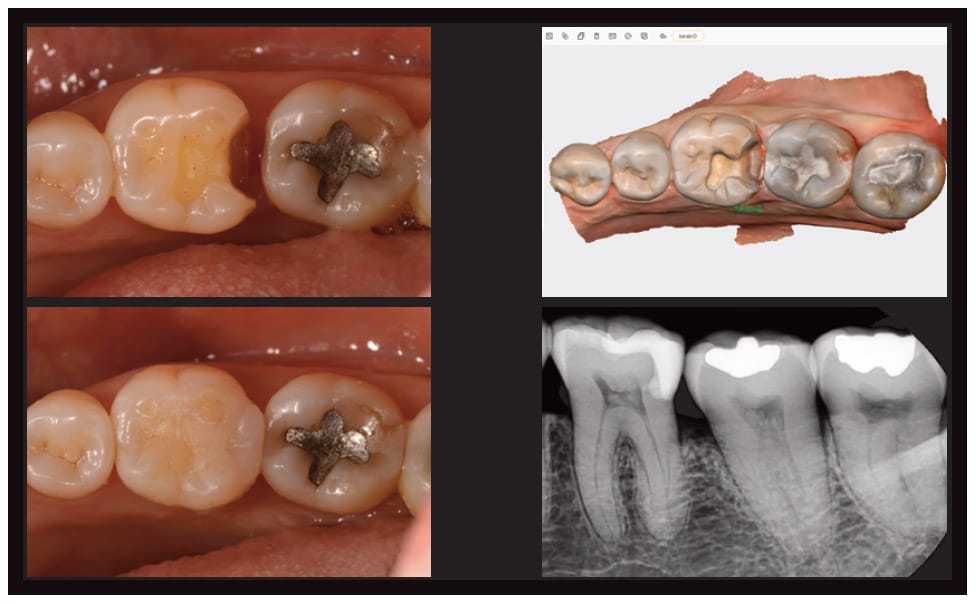
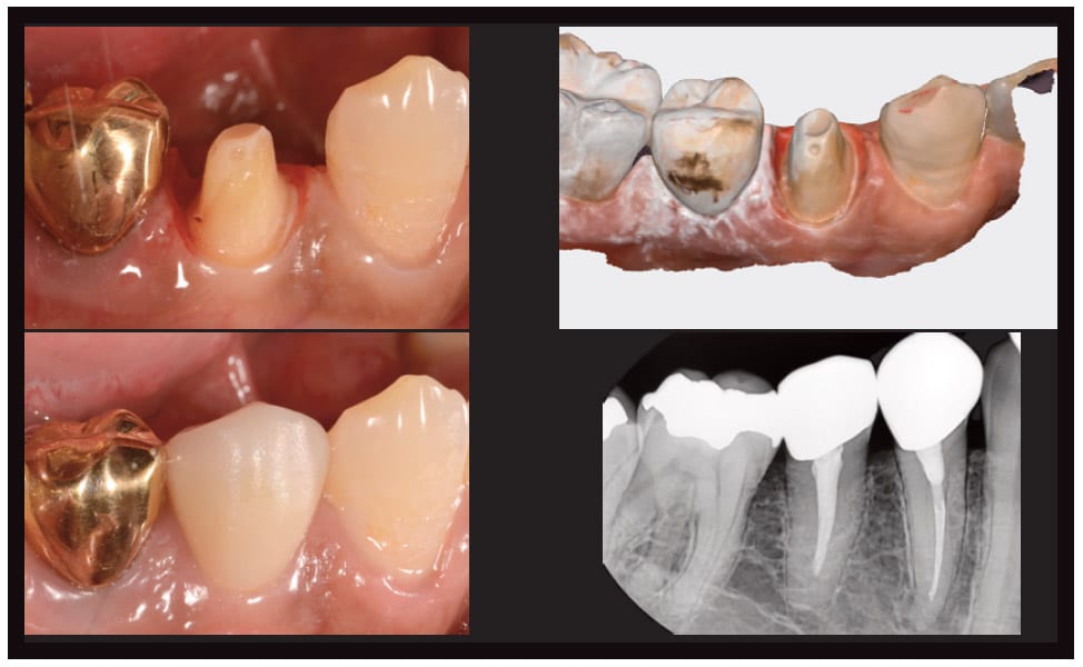
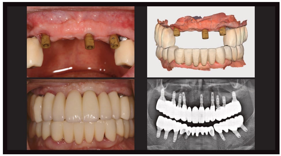
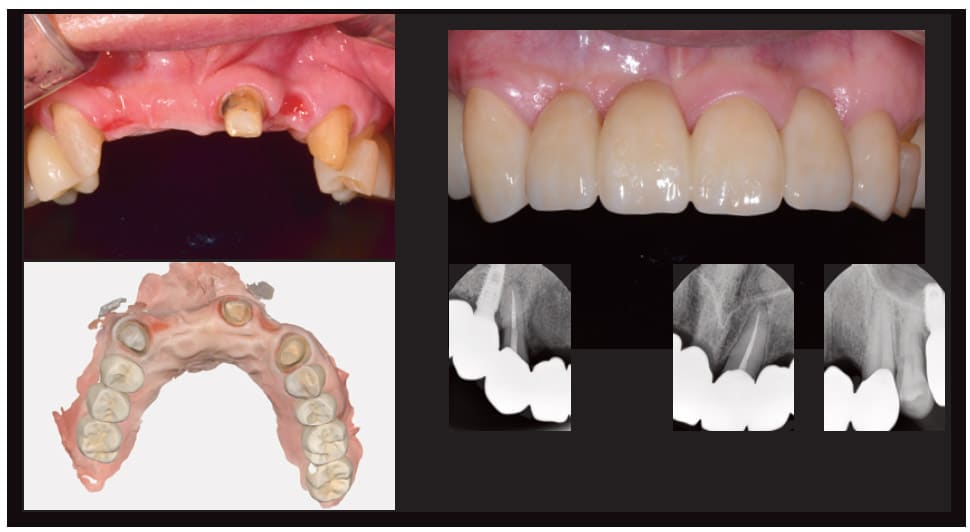
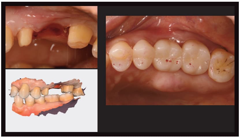
Many intraoral scanners are being used clinically as of 2022, and their reliability has increased significantly compared to just a few years ago. With the overall rapid advancement in performance, there are reports of digital scanning showing better marginal adaptation than conventional impressions in the case of a single restoration, as well as reports of significant improvement in the error range for complete arch scan data, which tended to fall short in accuracy compared to partial arches. My actual clinical experience is consistent with these findings. I’ve been using Medit’s i500 and i700 and obtaining satisfactory results through digital scanning when restoring natural teeth and implants (Figure 1 through Figure 4).
CONVENIENCE OF WIRELESS SCANNING
I had the opportunity to use the recently developed Medit i700 wireless and found it easier to apply intraorally and change scan positions. By sharing the screen of the scanner PC with the chair monitor, I was also able to conveniently move the scanner and scan without having to drag a scanner cart. I had no issue applying the i700 wireless to my clinical practice because there was no difference in scan data quality or frames per second compared to the wired device (Figure 5). I think I will use the i700 wireless more often in the future.
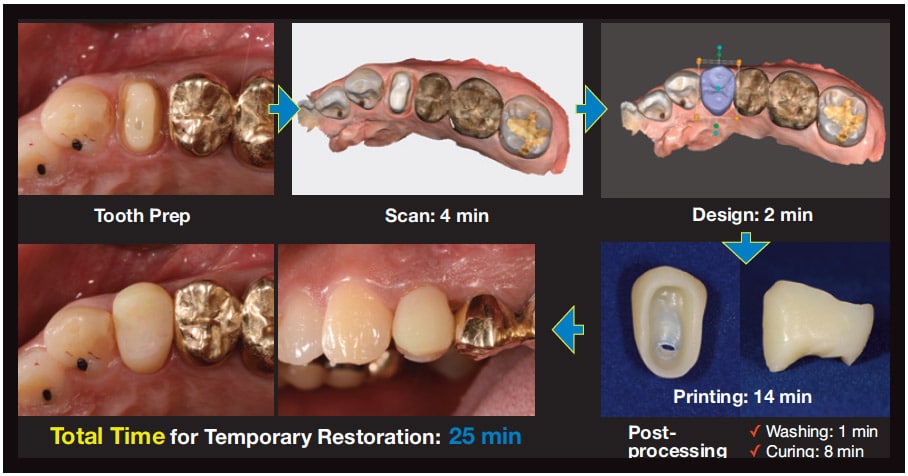
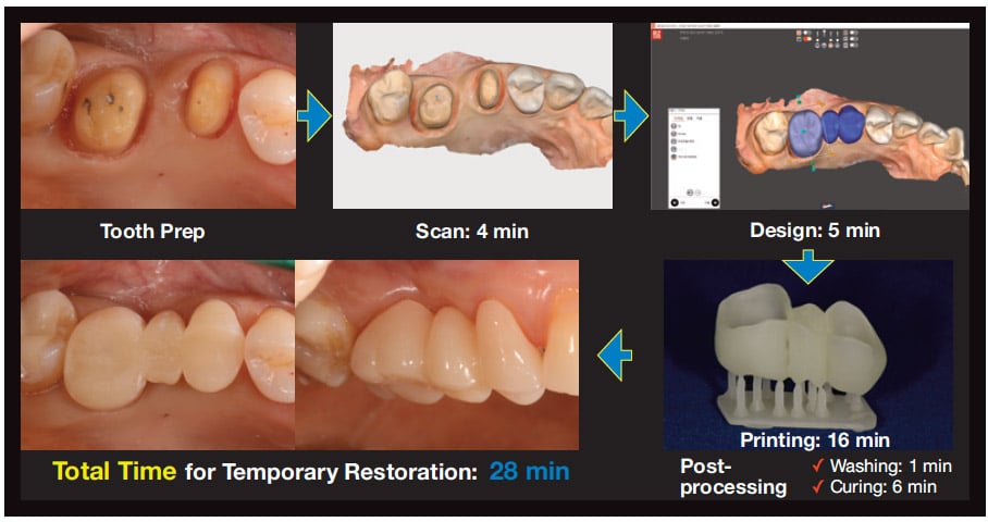
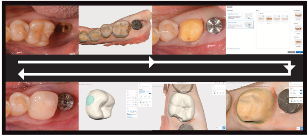
An intraoral scanner is attractive enough as an alternative to conventional impressions, but its greater advantage is the possibility of clinical expansion through scan data. My preferred clinical protocol is to digitally create temporary restorations. Intraoral scanning immediately creates a digital model, enabling a chairside digital workflow for provisional restorations. After preparations for zirconia crowns and intraoral scanning, I design crowns using a CAD program and create 3D-printed, or PMMA milled temporary restorations (Figure 6 and Figure 7).
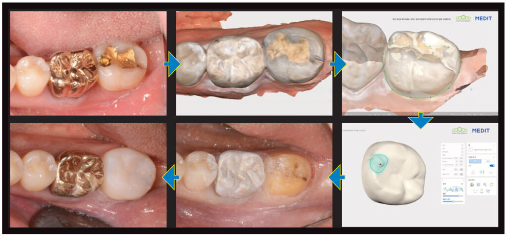
A CAD program is required to design the desired shape of restorations. In cases in which the shape of the original tooth before preparation can be used to create a temporary restoration, the Medit Temporaries App can be used to very simply design an eggshell temporary in the shape of the tooth before prep, or design a temporary restoration to fit the margin in the shape of a pre-prepared tooth after scanning (Figure 8 and Figure 9).
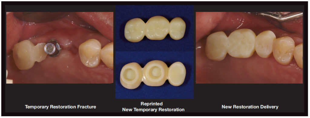
Digital production of temporary restorations is not only better than the analog method for efficient case management, but temporary restorations can also be reprinted anytime. When a temporary prosthesis breaks during use, as seen in Figure 9, it would have to be remade from scratch and would require a lot of time and effort. However, the digital workflow quickly solves this matter by reprinting temporaries when necessary (Figure 10).
DENTURE COPYING TECHNIQUE
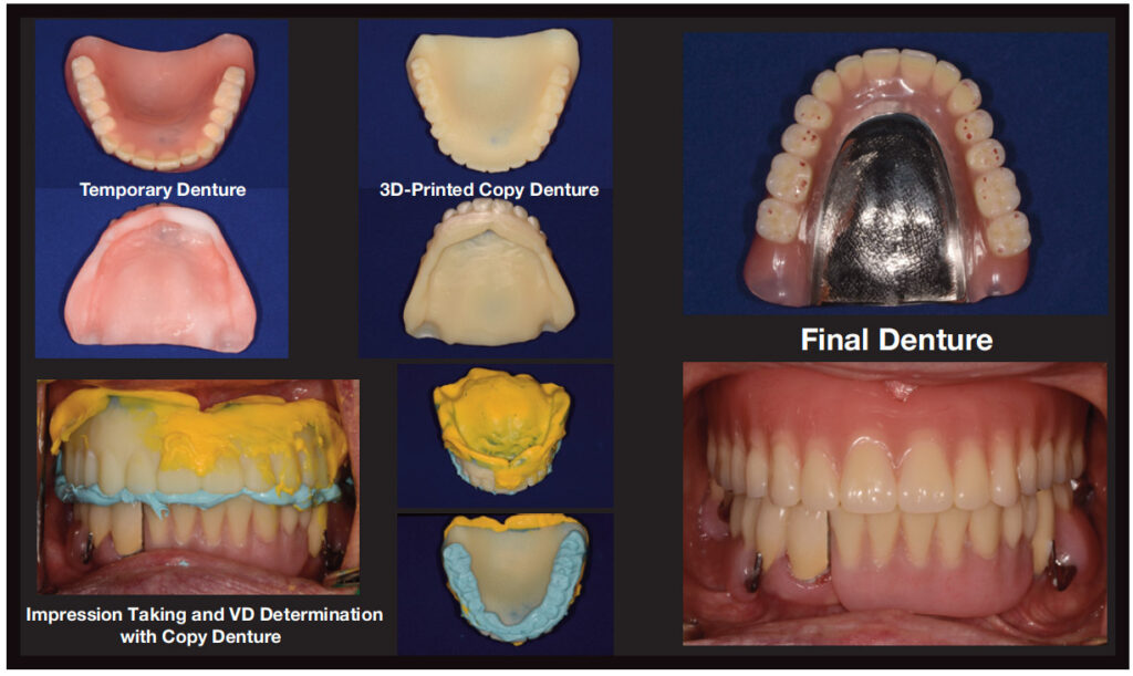
Copying dentures using intraoral scanners can also be a suitable protocol. Transferring the patient’s existing denture information to the copy denture significantly simplifies new denture production. In the case shown in Figure 11, we created a copy denture by scanning the temporary denture’s relined inner surface. It was 3D printed during the soft tissue healing phase after partial denture removal. Because this copy denture contains information of a suitable vertical dimension for the patient and has been border molded through several relinings, it can serve as both an individual tray and bite block.
By acquiring both impressions and bites using a copy denture, you can gather the required information to create a final denture at once. This makes denture creation much simpler than the usual production process of a complete denture.
SUMMARY
I’ve introduced some clinical applications using the Medit i700, but there are many more advantages of an intraoral scanner, such as preservation of data and better comparison views of oral changes. And intraoral scanner utilization will be significantly expanded through software updates in the future. I believe digital dentistry beginning with intraoral scanners is a workflow that one cannot and does not need to go against. I hope you can also experience the convenient and comfortable clinical changes I have experienced and am experiencing every day through digital dentistry.
MEDIT
From Decisions in Dentistry. July 2022;8(7)22,24.
