
Exploring Regenerative Approaches for Retrograde Peri-Implantitis
A novel case report highlights the potential of recombinant human platelet-derived growth factor with allograft material in managing retrograde peri-implantitis. While biofilm removal remains a challenge, this approach demonstrates promising outcomes in peri-implant bone regeneration and stability.
Retrograde peri-implantitis (RPI) presents a significant challenge in implant dentistry, often linked to prior endodontic complications and residual bacterial colonization. This case report explores a multidisciplinary approach incorporating antibiotics, mechanical debridement, and regenerative therapy using recombinant human platelet-derived growth factor with allograft material. The findings suggest a potential pathway for improving treatment outcomes, though further research is needed to establish standardized protocols.
The majority of RPI cases occur between 1 week and 4 years after implant placement with an increased prevalence for prior or adjacent endodontic complications.1,2 The treated RPI was diagnosed within that timeline. Residual bacteria may have been encapsulated in the bone even after debridement of the extraction socket and reactivated during implant bed preparation with subsequent colonization of the implant apex.3
Treatment was initiated with a course of antibiotics to address the active infection, localized swelling, and reduce the bacterial load prior to surgery. Despite its indications, the use of clindamycin is conflicting due to its adverse gastrointestinal effects and potential for significantly increased implant failure in penicillin-allergic patients.4
The decision to maintain the implant rested on the implant stability and defect anatomy. Regenerative flap therapy has been recommended for cases with peri-implant bone loss less than 50% and lack of implant mobility.5 In this case, despite the severe intrabony defect associated with the mesial root of the molar, extending to and including the apex of the implant, the lack of clinical mobility of the tooth and implant favored retention and subsequent periodontal regeneration. The patient was informed that the implant could still require removal and replacement over time.
A key step in the management of RPI is effective biofilm removal. A variety of strategies have been used for implant surface decontamination.6 Titanium scalers, rotary nylon brushes, and air polishing with glycine and erythritol powders have proven effective in eliminating calculus deposits and residual debris; however, undercuts, grooves, and porosities along roughened implant surfaces make it impossible to achieve a totally aseptic environment and introduce the potential for titanium dissolution into the surrounding tissues and microbial dysbiosis during instrumentation.7,8
Laser decontamination yielded positive results in animal models but its clinical use remains controversial with no true superiority to the aforementioned instruments in biofilm removal.9 Some chemotherapeutic agents, such as chlorhexidine, have more recently been shown to negatively alter the physicochemistry and cytocompatibility of titanium implants via the osteoblastic response, despite their long history as adjuncts intra- and post-operatively.10 At this time, there is no one mechanical or pharmacological agent that is superior in managing peri-implant lesions.8
Burnishing the implant surface with cotton pellets soaked in sterile saline has been reported to significantly reduce the level of pathogenic lipopolysaccharides and remove the peri-implant biofilm.8 Sterile saline is also cost effective with no cytotoxic effects to human osteoblasts.11
Tetracycline was used to further reduce the bacterial load along the implant body. Case reports in humans have shown that 50 mg/ml of tetracycline applied for 5 minutes after implantoplasty and followed by an autogenous bone graft or xenograft and membrane arrested disease progression and promoted radiographic bone fill of peri-implant defects.8,12 There is no definitive treatment for RPI and the evidence for most materials beyond sterile saline is conflicting.11,13 With this in mind, implant debridement with saline alone would have been sufficient for biofilm removal based on human and animal studies and should be considered in future applications of this protocol.8,10,11
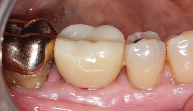
To the best of our knowledge, this is the first case report using rh-PDGF with an allograft material in the treatment of RPI. After 4 months, radiographs indicated adequate healing with marginal bone stability. The patient also had probing depths ≤ 4 mm with no bleeding on probing or suppuration. Studies have consistently found that true periodontal regeneration of cementum, periodontal ligament, and bone can be achieved using rh-PDGF with allograft, xenograft, and, to a lesser extent, β-TCP.14,15
In all cases, there was either a statistical improvement in bone regeneration or a strong trend toward bone gain in rh-PDGF-treated sites.14 Most recently, a case study reported the benefits of rh-PDGF on bone grafting procedures for severe ridge deficiencies in concomitance with periodontal attachment loss.15 In this case report, the bone replacement allograft was used as a scaffold and carrier for the rh-PDGF in a combined periodontal and peri-implant defect. The blend of cortical and cancellous bone particles in the allograft gives this product the structure of cortical chips, with the open scaffolding for bone to grow into offered by cancellous chips. The lack of osteogenic properties of these bone graft materials was overcome by the use of rh-PDGF.
This case report provides evidence for the potential use of rh-PDGF in challenging peri-implant defects; however, the treatment protocol described is not a standard clinical guideline. The use of chemotherapeutic agents in biofilm removal around implants is controversial.7,8,10 Further studies should be performed to validate these findings as they pertain to the surgical treatment of RPI.
References
- Wiedemann TG. A clinical approach to treatment of retrograde peri-implantitis. Compend Contin Educ Dent. 2021;42:170-175.
- Zhou W, Han C, Li D, Li Y, Song Y, Zhao Y. Endodontic treatment of teeth induces retrograde peri-implantitis. Clin Oral Implants Res. 2009;20:1326-1332.
- Marshall G, Canullo L, Logan RM, Rossi-Fedele G. Histopathological and microbiological findings associated with retrograde peri-implantitis of extra-radicular endodontic origin: a systematic and critical review. Int J Oral Maxillofac Surg. 2019;48:1475-1484.
- Zahra B, Nicholas B, Geoffrey R, Dina Z, Janal MN, Stuart F. Dental implant failure rates in patients with self-reported allergy to penicillin. Clin Implant Dent Relat Res. 2022;24:301-306.
- Sarmast ND, Wang HH, Sajadi AS, Munne AM, Angelov N. Nonsurgical endodontic treatment of necrotic teeth resolved apical lesions on adjacent implants with retrograde/apical peri-implantitis: a case series with 2-year follow-up. J Endod. 2019;45:645-650.
- Francis S, Iaculli F, Perrotti V, Piattelli A, Quaranta A. Titanium surface decontamination: a systematic review of in vitro comparative studies. Int J Oral Maxillofac Implants. 2022;37:76-84
- Kotsakis GA, Black R, Kum J, et al. Effect of implant cleaning on titanium particle dissolution and cytocompatibility. J Periodontol. 2021;92:580-591.
- Monje A, Amerio E, Cha JK, et al. Strategies for implant surface decontamination in peri-implantitis therapy. Int J Oral Implantol (Berl). 2022;15:213-248.
- Patil S, Bhandi S, Alzahrani KJ, et al. Efficacy of laser in re-osseointegration of dental implants-a systematic review. Lasers Med Sci. 2023;38:199.
- Kotsakis GA, Lan C, Barbosa J, et al. Antimicrobial agents used in the treatment of peri-implantitis alter the physicochemistry and cytocompatibility of titanium surfaces. J Periodontol. 2016;87:809-819.
- Brunello G, Becker K, Scotti L, Drescher D, Becker J, John G. The effects of three chlorhexidine-based mouthwashes on human osteoblast-like saos-2 cells. An in vitro study. Int J Mol Sci. 2021;22:9986.
- Reiser GM, Nevins M. The implant periapical lesion: etiology, prevention, and treatment. Compend Contin Educ Dent. 1995;16:768–772.
- Burdurlu MC, Dagasan VC, Tunc O, Guler N. Retrograde peri-implantitis: evaluation and treatment protocols of a rare lesion. Quintessence Int. 2021;52:112-121.
- Meghil MM, Mandil O, Nevins M, Saleh MHA, Wang HL. Histologic evidence of oral and periodontal regeneration using recombinant human platelet-derived growth factor. Medicina (Kaunas). 2023;59:676.
- Urban IA, Tattan M, Ravida A, Saleh MH, Tavelli L, Avila-Ortiz G. Simultaneous alveolar ridge augmentation and periodontal regenerative therapy leveraging recombinant human platelet-derived growth factor-bb (rhpdgf-bb): a case report. Int J Periodontics Restorative Dent. 2022;42:577-585.
This information originally appeared in Boeriu S, Hottel TL, Saltz AE, et al. A novel approach to treating retrograde peri-implantitis. Decisions in Dentistry. 2024;10(3):36-1.
