

Ridge Preservation of the Nonpreserved Socket
Appropriate surgical and prosthetic management of nonpreserved sockets following extractions will minimize alveolar bone loss, thus facilitating ridge preservation and implant success.
Appropriate surgical and prosthetic management of nonpreserved sockets following extractions will minimize alveolar bone loss, thus facilitating ridge preservation and implant success
In order to produce ideal functional and esthetic prostheses following dental implant therapy, sufficient alveolar bone volume and favorable architecture of the alveolar ridge are essential.1 It is well known that following tooth extraction, atrophy of the residual ridge occurs naturally,2,3 with the buccal bone plate resorbing considerably more during the healing process than the corresponding palatal/lingual bone walls.4,5 Alveolar bone loss can also occur as a result of iatrogenic trauma during tooth extraction.6 Schropp et al1 note the width of the alveolar ridge is reduced by 50% within the first year after extraction, with two-thirds of this reduction occurring within the first three months. In addition, loss of ridge height contributes to prosthetic instability, as the crest of the ridge approaches muscle attachments and mobile mucosa.7
Despite this knowledge, dental extractions are often performed without thought as to what may happen to the underlying alveolar bone, sometimes leading to a healed ridge that will not support an esthetic and functional prosthesis without significant bone grafting.6 When limited bone volume necessitates bone augmentation prior to implant placement, the potential need for additional surgical interventions should be discussed with the patient, as this increases the time and costs associated with treatment; it also raises the possibility of greater postoperative pain and limited implant survival.8
Ridge preservation includes any procedure that takes place at the time of, or shortly following, an extraction to minimize resorption of the ridge and maximize bone formation within the socket.9 Generally, the clinician uses a bone graft to fill the socket and places a membrane on top to prevent soft tissue ingrowth.10 When compared to extraction alone, this has been shown to limit the loss of hard tissue ridge width and provide a gain in hard tissue ridge height.11 If ridge preservation is not accomplished at the time of extraction, or should an atrophic ridge be present (for whatever reason) prior to implant placement, guided bone regeneration (GBR) can be used to augment bone in cases of vertical and/or horizontal defects.12 This technique consists of surgical placement of a cell occlusive membrane facing the bone surface, physically sealing off the skeletal site in need for regeneration, providing an environment permissive for recruitment and proliferation of osteoprogenitor cells, differentiation along the osteoblastic lineage, and expression of osteogenic activity.13 Survival rates of implants placed in areas augmented with GBR range from 92% to 100%.14
This clinical report highlights the treatment of a nonpreserved, edentulous space with a bone resorptive defect using GBR and subsequent implant placement.
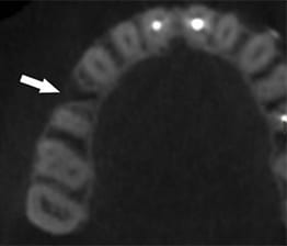
computed tomogram.
CLINICAL REPORT
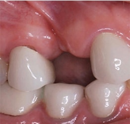
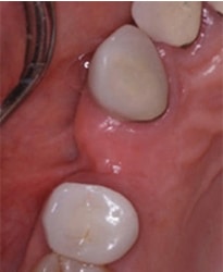
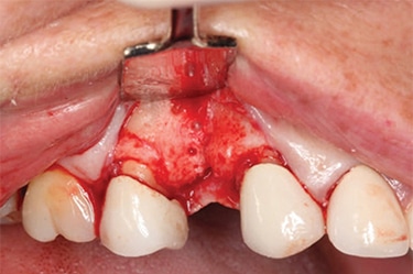
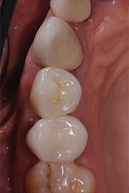
A 45-year-old female presented to the faculty practice clinic at the Dental College of Georgia at Augusta University with a missing maxillary right first premolar. The tooth had been extracted approximately seven months prior to the initial appointment. The patient presented with a noncontributory medical history. During the examination, it was determined the area of the extraction presented a high buccal frenum attachment (Figure 1) and loss of the buccal plate (Figure 2). Cone beam computed tomography (CBCT) was used to confirm the findings of the initial evaluation (Figure 3). The patient was referred to a periodontist for evaluation and treatment planning that included bone grafting, with simultaneous implant placement at the affected site.
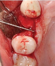
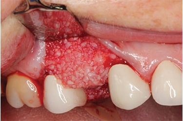
At the surgical appointment, local anesthesia was achieved with one carpule of lidocaine 2% with epinephrine 1:100,000 and one carpule of articaine 4% with epinephrine 1:100,000 to block the posterior superior alveolar, middle superior alveolar and palantine nerves. A midcrestal incision was made at the edentulous site and the team continued with intrasulcular incisions around adjacent teeth. The frenum attachment was relieved in the facial aspect, with a small mucosal perforation in the attachment area. A full-thickness mucoperiosteal flap was elevated. The tissue adherences were debrided. A significant bone resorption defect in the buccal aspect was noted, with a buccolingual width of 5 mm (Figure 4). A decision was made to postpone implant placement. The clinical team completed GBR with intracortical penetrations in the buccal aspect (Figure 5), demineralized freeze-dried bone allograft placement (Figure 6), and coverage with a 15×25-mm collagen membrane.
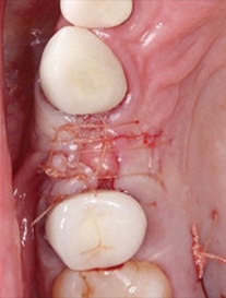
A connective tissue graft was obtained from the palatal tissue on the right side. The perforation in the frenum attachment area was corrected with the connective tissue graft. The donor site was sutured with a single interrupted 3-0 chromic suture technique, while the grafted site was sutured with horizontal mattress and single interrupted 3-0 chromic suture techniques. Hemostasis was achieved (Figure 7). At the follow-up appointment two weeks later, it was observed that primary tissue coverage was obtained, and the sutures were removed.
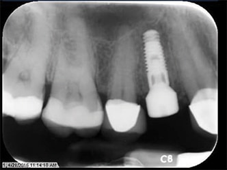
The patient was seen 90 days after the GBR appointment for implant placement surgery. Local anesthesia was achieved with one carpule of lidocaine 2% with 1:100,0000 epinephrine and one carpule of articaine 4% with 1:100,000 epinephrine, and one carpule of bupivacaine 0.5% with 1:100,000 epinephrine. A midcrestal incision was made to procure keratinized tissue in both the buccal and lingual aspects. The incision extended intrasulcularly to the distal of the right canine and mesial of the maxillary right second molar. A full-thickness mucoperiosteal flap was elevated, and a #4 round surgical diamond bur was used to mark the implant site. After using a pilot drill and taking a periapical radiograph, the osteotomy protocol was followed to prepare the bed for the implant. The site was copiously irrigated after the implant was placed and manually ratcheted. Type 2 bone was observed in site. Parallelism was obtained and verified with a periapical radiograph. Primary stability was achieved, and the healing collar was inserted using a hand-torque technique (Figure 8).
postoperative view.
A 1-mm fenestration in the buccal aspect was noticed, hence, a grafted cortical/cancellous mix was placed in the fenestration, and covered with a collagen wound dressing using the poncho technique.15 The team placed 4-0 vicryl sutures using the single interrupted and vertical mattress techniques. Hemostasis was achieved and gauze pressure packs were placed at the surgical site. The patient was seen at the two-week follow-up, at which time she reported no complications, so the sutures were removed.
The restorative phase was initiated three months after implant placement. After removal of the healing collar, the site was irrigated with 0.12% chlorhexidine gluconate. An implant level impression coping was screwed into place, and seating was verified by means of a periapical radiograph, followed by a full-arch, closed-tray impression using a hydrophilic vinyl polysiloxane material. The impression was visually checked for accuracy, the impression coping was removed and seated into the impression attached to an implant analog, and the healing collar was screwed back in place.
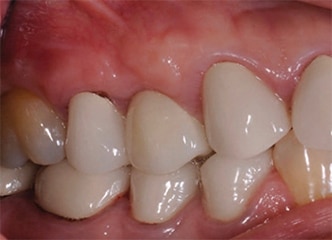
The clinical team chose a custom abutment with a buccal margin located 0.5 mm subgingivally to receive a cement-retained crown. The abutment was torqued into place at 30 Ncm. Full seating of the abutment was verified with a periapical radiograph. For protection against cement, the screw was covered with polytetrafluoroethylene tape. After try-in and verification of occlusion and interproximal contacts, a thin layer of resin modified glass ionomer cement was brushed on the intaglio surface of the crown and immediately placed over the custom abutment. Excess cement was thoroughly removed. A radiograph verified complete seating of the crown and that no excess cement remained around the margins. A follow-up appointment was scheduled at 13 months, at which time clinical and radiographic evaluation demonstrated stability of the implant and surrounding tissues. (Figure 9 and Figure 10).
DISCUSSION
Lack of planning and understanding of site preservation techniques immediately after an extraction could compromise the placement and long-term success of a dental implant. After CBCT evaluation by the team, it was clear the buccal bony defect in the alveolar ridge would require a graft. The minimum available bone width should be such that more than 1.0 mm of bone is present on either side of the implant buccolingually to keep the soft tissue levels stable. This is critical on the buccal aspect because any bone resorption and subsequent change in the position of the gingival margin may render nonesthetic results.16 After the GBR procedure was completed, the ridge measured at least 6 mm buccolingually, thus providing adequate bone thickness for a 3.7-mm-diameter implant.
A frenectomy was also performed prior to completion of GBR, not only to relieve pressure on the full-thickness mucoperiosteal flap and aid in coronal repositioning of the tissue, but also to prevent recession on the buccal aspect of the implant.17
The patient’s esthetics concerns and functional considerations were carefully evaluated when selecting the final prosthesis. The outcomes of screw-retained versus cemented single, implant-supported crowns have shown no significant difference in prosthetic complications.18 A custom titanium screw-retained implant abutment with a cemented implant-supported crown was selected over a screw-retained crown because there was adequate space (10 mm) between the edentulous ridge and opposing dentition that provided sufficient room for the prosthetic components.19
The effects of undetected excess cement on the peri-implant tissue are well known, although there are a few studies confirming their effects.20,21 Compared to methacrylate cements, the use of temporary luting material for the cementation of restorations on implants has fewer clinical risks.22 Temporary luting cements are less retentive than other cements,23 however, which could lead to crown dislodgement and (unnecessary) additional chairtime. In this case, the use of a radiopaque resin modified glass ionomer allowed the team to detect any excess cement radiographically. In addition, the location of the abutment margin at no more than 0.5 mm subgingivally facilitated cement removal — which is a key factor in preventing peri-implant inflammation and peri-implant bone loss.
SUMMARY
Atraumatic extractions and ridge preservation are important aspects in ensuring the long-term success of implant therapy. When providers do not consider these factors, bone loss and/or ridge defects can occur that may compromise the esthetic outcome of any restorative plan. In these cases, bone regeneration procedures are highly recommended prior to implant placement.24 Ultimately, adopting a multidisciplinary approach aids in the long-term outcome and prognosis of the final restoration.
Acknowledgement:
The authors thank William Brackett, DDS, MS, for his guidance and contributions to this manuscript.
KEY TAKEAWAYS
- Lack of planning and understanding of site preservation techniques immediately after a tooth extraction could compromise the long-term success of implant therapy.
- Sufficient alveolar bone volume and favorable architecture of the alveolar ridge are needed to produce esthetic and highly functional implant prostheses.1
- Atrophy of the residual ridge occurs naturally following tooth extraction,2,3 and alveolar bone loss can also occur as a result of iatrogenic trauma during these procedures.6
- Ridge preservation includes any therapy that takes place at the time of, or shortly following, an extraction to minimize resorption of the ridge and maximize bone formation within the socket.9
- Generally, the clinician uses a bone graft to fill the socket and places a membrane on top to prevent soft tissue ingrowth.10
- Atraumatic extractions and ridge preservation are important considerations during implant therapy.
- Adopting a multidisciplinary approach aids in the long-term outcome and prognosis of the final restoration.
REFERENCES
- Schropp L, Wenzel A, Kostopoulos L, Karring T. Bone healing and soft tissue contour changes following single-tooth extraction: a clinical and radiographic 12-month prospective study. Int J Periodontics Restorative Dent. 2003;23:313–323.
- Atwood DA. Reduction of residual ridges: a major oral disease entity. J Prosthet Dent. 1971;26:266–279.
- Pietrokovski J, Massler M. Alveolar ridge resorption following tooth extraction. J Prosthet Dent. 1967;17:21–27.
- Trombelli L, Farina R, Marzola A, Bozzi L, Liljenberg B, Lindhe J. Modeling and remodeling of human extraction sockets. J Clin Periodontol. 2008;35:630–639.
- Araujo MG, Lindhe J. Dimensional ridge alterations following tooth extraction. An experimental study in the dog. J Clin Periodontol. 2005;32:212–218.
- Irinakis T, Tabesh M. Preserving the socket dimensions with bone grafting in single sites: an esthetic surgical approach when planning delayed implant placement. J Oral Implantol. 2007;33:156–163.
- Bartee BK. Extraction site reconstruction for alveolar ridge preservation. Part 1: rationale and materials selection. J Oral Implantol. 2001;27:187–193.
- Zitzmann NU, Krastl G, Hecker H, Walter C, Waltimo T, Weiger R. Strategic considerations in treatment planning: deciding when to treat, extract, or replace a questionable tooth. J Prosthet Dent. 2010;104:80–91.
- Darby I, Chen S, De Poi R. Ridge preservation: what is it and when should it be considered. Aust Dent J. 2008;53:11–21.
- Apostolopoulos P, Darby I. Retrospective success and survival rates of dental implants placed after a ridge preservation procedure. Clin Oral Implants Res. 2017;28:461–468.
- Iasella JM, Greenwell H, Miller RL, et al. Ridge preservation with freeze-dried bone allograft and a collagen membrane compared to extraction alone for implant site development: a clinical and histologic study in humans. J Periodontol. 2003;74:990–999.
- Clementini M, Morlupi A, Canullo L, Agrestini C, Barlattani A. Success rate of dental implants inserted in horizontal and vertical guided bone regenerated areas: a systematic review. Int J Oral Maxillofac Surg. 2012;41:847–852.
- Retzepi M, Donos N. Guided bone regeneration: biological principle and therapeutic applications. Clin Oral Implants Res. 2010;21:567–576.
- Chiapasco M, Zaniboni M, Boisco M. Augmentation procedures for the rehabilitation of deficient edentulous ridges with oral implants. Clin Oral Implants Res. 2006;17(Suppl 2):136–159.
- Hoexter DL, Epstein SB. Poncho graft. Oral Implantol. 1974;5:210–218.
- Belser UC, Bernard JP, Buser D. Implant-supported restorations in the anterior region: Prosthetic considerations. Pract Periodontics Aesthet Dent. 1996;8:875–883.
- Fu JH, Su CY, Wang HL. Esthetic soft tissue management for teeth and implants. J Evid Based Dent Pract. 2012;12(Suppl 3):129–142.
- Amorfini L, Storelli S, Mosca D, Scanferla M, Romeo E. Comparison of cemented vs screw-retained, customized computer-aided design/computer-assisted manufacture zirconia abutments for esthetically located single-tooth implants: a 10-year randomized prospective study. Int J Prosthodont. 2018;31:359–366.
- Ganz SD. An implant restoration as the primary treatment alternative replacing a maxillary first bicuspid tooth. Implant Soc. 1992;3:2–5.
- Korsh M, Obst U, Walther W. Cement-associated peri-implatitis: a retrospective clinical observational study of fixed implant-supported restorations using a methacrylate cement. Clin Oral Implants Res. 2014;25:797–802.
- Wilson TG Jr. The positive relationship between excess cement and peri-implant disease: a prospective clinical endoscopic study. J Periodontol. 2009;80:1388–1392.
- Korsh M, Walther W. Peri-implantitis associated with type of cement: a retrospective analysis of different types of cement and their clinical correlation to the peri-implant tissue. Clin Oral Implant Dent Relat Res. 2015;17(Suppl):e434–e443.
- Grag P, Gupta G, Prithviraj DR, Pujari M. Retentiveness of various luting agents used with implant-supported protheses: a preliminary in vitro study. Int J Prosthodont. 2013;26:82–84.
- Byrne G. Socket preservation of implant sites. J Am Dent Assoc. 2012;143:1139–1140.
The authors have no commercial conflicts of interest to disclose
Featured image by SHCHERBAK VOLODYMYR/ISTOCK/GETTY IMAGES PLUS
From Decisions in Dentistry. December 2018;4(12):12—14,17.
