
Dental Laser Applications in Pediatrics
With indications ranging from soft tissue surgery to caries preparation, dental lasers can help clinicians maintain optimal oral health in this patient population.
With indications ranging from soft tissue surgery to caries preparation, dental lasers can help clinicians maintain optimal oral health in this patient population
PURCHASE COURSE
This course was published in the July 2018 issue and expires July 2021. The author has no commercial conflicts of interest to disclose. This 2 credit hour self-study activity is electronically mediated.
This course was published in the July 2018 issue and expires July 2021. The author has no commercial conflicts of interest to disclose. This 2 credit hour self-study activity is electronically mediated.
EDUCATIONAL OBJECTIVES
After reading this course, the participant should be able to:
- Explain laser development and technologies, operating modes, and related safety considerations.
- Describe common applications for dental lasers in pediatric populations.
- Cite examples of soft tissue laser procedures suitable for children and adolescents.
Einstein first described laser function in 1917, although the actual technology was not developed until the 1960s.1 Laser energy is created by intensified light, which is generated by stimulating a synthetic medium inside a light chamber. Lasers are typically named after the active medium that generates the photons.1,2 The most commonly used dental laser technologies include carbon dioxide (CO2), erbium-doped yttrium aluminum garnet (Er:YAG), neodymium-doped yttrium aluminum garnet (Nd:YAG), neodymium-doped yttrium aluminum perovskite (Nd:YAP), holmium yttrium aluminum garnet (HO:YAG), gallium arsenide (diode), erbium chromium-doped yttrium scandium gallium garnet (Er-Cr:YSGG), and argon lasers. Indicated for both adult and pediatric patients, medical and dental lasers have variable wavelengths, and operate in continuous-wave, pulsed or running-pulsed modes.3
Laser applications for medical science began in the 1970s,4 and the first application in dentistry was as a replacement for scalpels and cautery instruments in oral soft tissue surgery.3 Applications of laser technologies depend on the absorption coefficients of the tissues at various wavelengths.1 Specifically, lasers in the visible and near-infrared spectrum absorbed by hemoglobin and melanin are used to treat soft tissue pathologies. Erbium-family lasers are absorbed by water in gingiva, mucosa, and hydroxyapatite of enamel, and have both hard and soft tissue applications. CO2 lasers are absorbed by water in mucosa and gingiva, and are primarily used for gingival surgery. However, studies also show absorption by hydroxyapatite, and CO2 lasers may have use in preventing acidic dissolution of enamel.4
The American Academy of Pediatric Dentistry (AAPD) has developed a policy regarding the use of lasers for treating children and adolescents.5 This modality and other approaches help oral health professionals work with patients and parents/caregivers in maintaining patients’ optimal oral health. The AAPD policy paper recognizes that this goal may be achieved by using lasers as an alternative and complimentary instrument to perform selected procedures.
Safety is imperative when utilizing lasers in clinical practice. Protective eyewear is essential for the patient, provider, dental team members, and others observing the procedure. In addition, there is risk of disease transmission via viral particles contained in laser-generated aerosols. Caution should be used for immunocompromised patients, and pharmacological therapy may be treatment of choice in these cases.5–8 Sufficient operator training, appropriate laser selection, and effective safety measures are essential when providing laser treatment to pediatric populations.
DIAGNOSTIC APPLICATIONS
It is the goal of oral health professionals to establish an accurate diagnosis of caries. Up to 25% of children and 50% of adolescents will experience caries in their permanent teeth, and rates of caries in primary teeth are even greater.9 Laser technology has been developed and studied as an adjunct to clinical and radiographic caries diagnosis. Detection of demineralization of tooth structure has been described with the use of dental lasers. Analysis of the light reflected from occlusal surfaces of primary and permanent teeth is used to detect demineralization and changes in tooth structure. However, sealants, composite resin, residual toothpaste and debris may alter readings and lead to false positives.2,4,5,8,10 In light of the possibility of false positive readings, providers should perform accurate and serial assessment of caries activity. Laser detection of caries could reduce radiation exposure to children by extending the interval between radiographs. That noted, given the incidence of false positives, diagnosis using laser technology is recommended as an adjunct to traditional radiographic and clinical analysis.
SOFT TISSUE APPLICATIONS
There are many applications for pediatric soft tissue laser procedures in oral surgery, oral pathology, periodontology and orthodontics — including frenectomies, frenotomies, operculectomies, gingivectomies, and treatment of aphthous and herpetic lesions.5–7,11
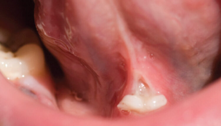
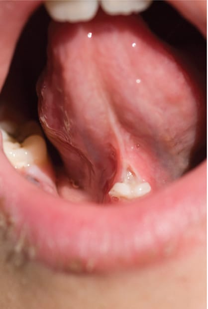
Ankyloglossia of the infant is a common and well-studied condition in which the attachment of the lingual frenum is at the tip of the tongue. This is most common in boys, with a reported incidence of 4% to 5%.11,12 Ankyloglossia and restriction of tongue movement can result in poor breastfeeding and maternal nipple pain. Research shows that frenotomy can reduce maternal nipple pain, although the effect on breastfeeding quality has not been consistently proven.13 Frenotomy involves administering local anesthesia and using two instruments — such as a mosquito or hemostat — in a V shape to protect structures in the sublingual area, including the lingual nerve and Wharton’s duct.14 A scalpel, laser or electrosurgical knife is used to make a V-shaped incision separating the attachment. Advantages of using a laser include the specific interaction of the laser with treated tissue, rapid hemostasis, reduced postoperative discomfort and fast wound healing.11 The laser also has a decontaminating and antibacterial effect.11
Laser labial frenectomy is completed in a similar fashion, but can also include surgical repositioning of the frenum attachment. While indications for labial frenectomy are varied, this procedure can improve access and cleansability of the facial surfaces of the young child’s incisors, reduce traction on the marginal gingiva, aid in orthodontic treatment of a persistent midline diastema, and modify labial or lingual anatomic issues in a multidisciplinary approach to speech development.15 Advantages of using a laser in frenectomy or frenotomy include reduced operating time, decreased use of local anesthetic and faster healing.
Removal of benign, diseased oral soft tissue, such as mucocele, gingival fibroma, and pyogenic granuloma, can also be achieved with a diode or Nd:YAG laser. Case reports also indicate successful use of Er-Cr:YSGG lasers for these procedures.16 Aphthous ulcers and herpetic ulcers may be treated with laser therapy, resulting in reduced healing time — although more high-quality studies are needed to determine effectiveness and treatment protocols.17
Periodontal disease is found in a smaller percentage of pediatric patients than adults; nonetheless, teenagers and children present with various periodontal conditions. Some of the most common laser procedures performed in a pediatric population include gingivectomy, removal of operculum, or exposure of a submerged tooth for placement of an orthodontic bracket.6 Although laser decontamination of periodontal pockets in adults has been studied with varying results, there is little research in pediatric populations. At this time, laser therapy is best considered as an adjunct to traditional periodontal treatment.18
Additionally, evolving fields in medicine and dentistry involve photobiomodulation (PBM) and low-level laser therapy (LLLT). In PBM, lasers are used to initiate a photochemical effect on a cellular level, without thermal or ablative characteristics. The majority of studies use diode lasers and attribute positive clinical changes to stimulation of mitochondrial cytochromes, eliciting increased cellular metabolism and improved cell viability.19 While more studies are needed, there is sufficient literature to support PBM in the treatment of oral mucositis — which is of interest to clinicians who manage patients undergoing radiation or chemotherapy.
HARD TISSUE APPLICATIONS
Comfortable, minimally invasive dentistry is an important goal in pediatric care. Tooth preparation using lasers represents an opportunity to provide comfortable and conservative restorative treatment.20 In this capacity, Er:YAG technology is most commonly used for hard tissue applications. In caries preparation, the erbium laser family has the advantage of providing some analgesic effect on the tooth structure, as well as reducing bacterial content at the prepared site.10 Laser interaction and effectiveness in removing hard tissue depends on tooth composition, including the water and mineral content of hydroxyapatite. As such, primary teeth often require a lower power setting than permanent teeth.21 In general, as the operator moves the laser closer to the pulp tissue, power settings are decreased, with the goal of comfortable and conservative caries preparation. In Class II preparations, a matrix may be recommended to protect against laser etching of adjacent tooth structure.10 Adherence to manufacturer guidelines for the particular laser in use is important for effective and efficient removal of hard tooth structure.
Although operating time is reported to be longer with laser preparation, the requirement for local anesthesia is reduced due to the laser’s analgesic effect.22 If this helps ease anxiety over injections, it can be especially important for behavior management in children and adolescents. Additionally, a laser does not produce the same noise and vibration as a dental handpiece, which has been discussed as a potential source of anxiety in pediatric patients.5 Because lasers are primarily end cutting, a slow-speed instrument may be needed for preparation refinement following laser use.23
Bond strength to restorations is paramount when considering preparation techniques. While composite bond strength supports more conservative caries preparations, Olivi et al20 reported decreased bond strength with laser preparation and glass ionomer compared to traditional handpiece-based restorative techniques. Other studies suggest no differences in microleakage for composite restorations, but do not consistently address restoration retention in a clinical setting.22 More studies, especially in pediatric populations, are necessary to determine best practices for material selection and long-term success in primary and permanent teeth.
Erbium lasers can also be used for enamel etching prior to placing sealants; however, this technique has not been proven to improve sealant bond strength or retention. Most current studies indicate that microleakage is minimal with phosphoric acid etching; in addition, there is no clinically significant difference in sealant retention between laser and phosphoric acid etching.24,25
PULPAL THERAPY
The American Association of Endodontists published a position paper on the use of lasers.26 In summary, there are advantages and disadvantages to laser use in endodontic therapy, and root disinfection is likely more useful than root preparation. Advantages in root disinfection include photodynamic therapy to kill microorganisms, as well as photon-induced acoustic streaming to distribute disinfecting solutions across root canal systems. Disadvantages include the curved nature of root canal systems, heat generation, and the lack of clinical evidence supporting the long-term success of laser use. That noted, the U.S. Food and Drug Administration (FDA) has approved diode lasers for pulpotomies and apicoectomies.
In pediatric dentistry, pulpotomy is a common vital pulp procedure that seeks to maintain a tooth by removing infected coronal pulp tissue and preserving radicular pulp tissue.6,26–28 Laser pulpotomy has been studied as a replacement for other commonly used medicaments, such as formocresol, mineral trioxide aggregate (MTA), ferric sulfate, calcium hydroxide and sodium hypochlorite. Laser use is often sought out due to concerns regarding unnecessary exposure to various medicaments. The diode is the most commonly used (and FDA approved) laser for pulpotomies, although CO2, Er:YAG and Nd:YAG laser use has also been reported.28 Overall success rates for formocresol, MTA, ferric sulfate, sodium hypochlorite and lasers are approximately 80% at 24 months. When compared to other medicaments used in pulpotomy of primary teeth, in most studies, the laser showed no advantage, while MTA or formocresol had the highest success rates. In light of available studies, and based on low-quality evidence, the AAPD guidelines for vital pulp therapy conditionally recommend lasers for pulpotomies in primary teeth.27
DENTAL TRAUMA AND ORAL SURGERY
Dental trauma is common in childhood, as reports indicate that 20% of children injure their primary teeth and 15% injure permanent dentition.29 Hence, pediatric dentists and general practitioners often assess and treat traumatic injuries. A thorough history and examination — as well as ruling out more serious conditions, such as a closed head injury — are essential. Laser treatment of pulp tissue involved in complicated crown fractures has been studied (primarily with case reports in pediatric populations). Similar to research for laser pulpotomy and pulpectomy, when compared to other medicaments, evidence regarding laser use for pulp treatment is either insufficient or inconclusive.27—29 However, injuries — such as luxation, subluxation, or avulsion — may benefit from decontamination of the periodontal pocket or tooth socket to promote attachment healing. This decontamination can be achieved using a diode or Nd:YAG laser.29 Novel research in LLLT has shown beneficial results for soft tissue, mucosal and gingival healing that may apply to dental trauma. Research in oral surgery and bone healing also suggests that LLLT may facilitate healing, reduce inflammation and offer superior pain management.30,31 More research in these areas — and in pediatric populations — is necessary to establish treatment protocols and evaluate effectiveness.
ORTHODONTIC TREATMENT
Treatment-associated pain and discomfort in orthodontic care is commonly noted in children and adolescents. Pain may present as discomfort or hypersensitivity of affected teeth. Reduction of pain associated with these procedures has been described using LLLT, with some studies showing an analgesic effect lasting up to 72 hours after placement of light archwires and elastomeric separators.32 As noted with many studies regarding LLLT, however, there is a lack of randomized controlled trials and high-quality evidence. Until protocols are established, clinical judgement should be used when prescribing LLLT.
SUMMARY
Laser therapy represents a novel and exciting complementary treatment modality for oral health professionals. With applications ranging from soft tissue surgery to caries preparation, the potential advantages include faster healing after trauma and surgery, and reduced pain following procedures. As with other clinical techniques, training and safety are of the utmost concern when treating pediatric patients. That said, a reduced need for anesthetic during tooth preparation, and the potential for improved healing in frenotomies, frenectomies and minor soft tissue surgeries represent valid reasons to consider lasers as a viable approach to pediatric treatment.
REFERENCES
- Martens LC. Laser physics and a review of laser applications in dentistry for children. Eur Arch Paediatr Dent. 2011;12:61–67.
- Stabholz A, Zeltser R, Sela M, et al. The use of lasers in dentistry: principles of operation and clinical applications. Compend Contin Educ Dent. 2003;24:935–949.
- Nazemisalman B, Farsadeghi M, Sokhansanj M. Types of lasers and their applications in pediatric dentistry. J Lasers Med Sci. 2015;6:96–101.
- Caprioglio C, Olivi G, Genovese MD. Paediatric laser dentistry. Part 1: general introduction. Eur J Paediatr Dent. 2017;18:80–82.
- Policy on the use of lasers for pediatric dental patients. Pediatr Dent. 2016;38:84–86.
- Olivi G, Caprioglio C, Olivi M, Genovese MD. Paediatric laser dentistry. Part 4: soft tissue laser applications. Eur J Paediatr Dent. 2017;18:332–334.
- Kumar G, Rehman F, Chaturvedy V. Soft tissue applications of Er,Cr:YSGG laser in pediatric dentistry. Int J Clin Pediatr Dent. 2017;10:188–192.
- Kotlow L. Lasers and pediatric dental care. Gen Dent. 2008;56:618–627.
- Wright JT, Tampi MP, Graham L, et al. Sealants for preventing and arresting pit-and-fissure occlusal caries in primary and permanent molars. Pediatr Dent. 2016;38:282–308.
- Kotlow LA. Lasers in pediatric dentistry. Dent Clin North Am. 2004;48:889–922.
- Crippa R, Paglia M, Ferrante F, Ottonello A, Angiero F. Tongue-tie assessment: clinical aspects and a new diode laser technique for its management. Eur J Paediatr Dent. 2016;17:220–222.
- Power RF, Murphy JF. Tongue-tie and frenotomy in infants with breastfeeding difficulties: achieving a balance. Arch Dis Child. 2015;100:489–494.
- O‘Shea JE, Foster JP, O‘Donnell CP, et al. Frenotomy for tongue-tie in newborn infants. Cochrane Database Syst Rev. 2017;3:CD011065.
- Kotlow L. Diagnosis and treatment of ankyloglossia and tied maxillary fraenum in infants using Er:YAG and 1064 diode lasers. Eur Arch Paediatr Dent. 2011;12:106–112.
- Olivi M, Genovese MD, Olivi G. Laser labial frenectomy: a simplified and predictable technique. Retrospective clinical study. Eur J Paediatr Dent. 2018;19:56–60.
- Kumar G, Rehman F, Chaturvedy V. soft tissue applications of Er,Cr:YSGG laser in pediatric dentistry. Int J Clin Pediatr Dent. 2017;10:188–192.
- Han M, Fang H, Li Q, Cao Y, Xia R, Zhang ZH. Effectiveness of laser therapy in the management of recurrent aphthous stomatitis: a systematic review. Scientifica (Cairo). 2016;2016:9062430.
- Meimandi M, Talebi Ardakani MR, Nejad AE, Yousefnejad P, Saebi K, Tayeed MH. The effect of photodynamic therapy in the treatment of chronic periodontitis: a review of literature. J Lasers Med Sci. 2017;8:S11.
- Pandeshwar P, Roa MD, Das R, Shastry SP, Kaul R, Srinivasreddy MB. Photobiomodulation in oral medicine: a review. J Investig Clin Dent. 2016;7:114–126.
- Olivi G, Caprioglio C, Olivi M, Genovese MD. Paediatric laser dentistry. Part 2: hard tissue laser applications. Eur J Paediatr Dent. 2017;18:163–166.
- Zhegova G, Rashkova M, Rocca JP. Minimally invasive treatment of dental caries in primary teeth using an Er:YAG laser. Laser Ther. 2014;23:249–254.
- Tao S, Li L, Yuan H, et al. Erbium laser technology vs traditional drilling for caries removal: a systematic review with meta-analysis. J Evid Based Dent Pract. 2017;17:324–334.
- Montedori A, Abraha I, Orso M, D‘Errico PG, Pagano S, Lombardo G. Lasers for caries removal in deciduous and permanent teeth. Cochrane Database Syst Rev. 2016;9:CD010229.
- Fumes AC, Longo DL, De Rossi A, et al. Microleakage of sealants after phosphoric acid, Er: YAG laser and air abrasion enamel conditioning: systematic review and meta-analysis. J Clin Pediatr Dent. 2017;41:167–172.
- Kumar G, Dhillon JK, Rehman F. A comparative evaluation of retention of pit and fissure sealants placed with conventional acid etching and Er,Cr:YSGG laser etching: a randomised controlled trial. Laser Ther. 2016;25:291–298.
- American Association of Endodontists. Clinical Guidelines & Position Statements. Available at: www.aae.org/specialty/clinical-resources/guidelines-position-statements/. Accessed May 24, 2018.
- Dhar V, Marghalani AA, Crystal YO, et al. Use of vital pulp therapies in primary teeth with deep caries lesions. Pediatr Dent. 2017;39:173–186.
- De Coster P, Rajasekharan S, Martens L. Laser-assisted pulpotomy in primary teeth: a systematic review. Int J Paediatr Dent. 2013;23:389–399.
- Caprioglio C, Olivi G, Genovese MD, Vitale MC. Paediatric laser dentistry. Part 3: dental trauma. Eur J Paediatr Dent. 2017;18:247–250.
- Carroll JD, Milward MR, Cooper PR, Hadis M, Palin WM. Developments in low level light therapy (LLLT) for dentistry. Dent Mater. 2014;30:465–475.
- Noba C, Mello-Moura AC, Gimenez T, Tedesco TK, Moura-Netto C. Laser for bone healing after oral surgery: systematic review. Lasers Med Sci. 2018;33:667–674.
- Deana NF, Zaror C, Sandoval P, Alves N. Effectiveness of low-level laser therapy in reducing orthodontic pain: a systematic review and meta-analysis. Pain Res Manag. 2017;2017:8560652.
From Decisions in Dentistry. July 2018;4(7):28–31.

