

Conservative Restoration of a Maxillary Central Incisor With Severe Crown Dilaceration
Treating a maxillary crown dilaceration often involves multidisciplinary care, including orthodontic, surgical, endodontic and restorative therapies.
Treating a maxillary crown dilaceration often involves multidisciplinary care, including orthodontic, surgical, endodontic and restorative therapies
Dilaceration refers to a sharp angulation between the root and crown of a formed tooth.1 Trauma is a common cause of root dilacerations, particularly in the maxillary anterior region. While dilaceration can occur anywhere along the length of the tooth, crown dilaceration constitutes 3% of all traumatic dental injuries, and is more common in maxillary anterior teeth compared with mandibular anterior dentition.2–7
The treatment of maxillary crown dilaceration is complicated and often involves multidisciplinary care.8 Depending on the clinical status of a dilacerated tooth, various treatment modalities may be necessary, including orthodontic, surgical, endodontic and restorative therapies.8–13 Although treatment might involve different disciplines, restorative interventions are necessary in most crown dilaceration cases to provide acceptable functional and esthetic outcomes.14–18 Furthermore, while replacing missing teeth or teeth with poor prognoses with dental implants is a common and predictable treatment, placing implants in young adults who are still growing is a challenge, especially in the esthetic zone.19–22
This clinical report describes a conservative and predictable treatment to restore function and esthetics in a maxillary central incisor with severe crown dilaceration.
CLINICAL REPORT
An 18-year-old male was referred by the Texas A&M College of Dentistry’s Orthodontic Department for an extraction and replacement of a maxillary left central incisor (tooth #9) with a dental implant. A comprehensive examination and all necessary radiographs were completed. The patient had a low caries risk, with no active caries or signs of periodontal disease. A comprehensive medical history revealed no contraindication for elective dental treatment. Reviewing the cone beam computed tomography report revealed a 90-degree root dilaceration of tooth #9.

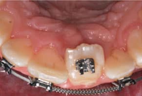
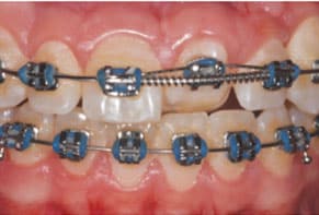
After considering the patient’s skeletal maturation index at the time, a surgical consult with a periodontist was also completed due to concerns over tooth extraction and implant placement, as this patient will continue to grow.23 Following a complete clinical evaluation and multidisciplinary discussion, various treatment options were reviewed with the patient. These included restoring the existing tooth (with or without root canal treatment), post core and crown, replacing the existing tooth with an implant-supported crown (considering the consequences of growing), resin bonded fixed dental prosthesis (FDP), conventional tooth-supported FDP, or removable prosthesis (Figure 1 and Figure 2).
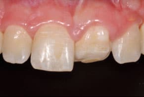
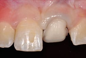
The final treatment plan was to use a partial or full-coverage all-ceramic crown, noting the potential risk of pulpal exposure during tooth preparation. This treatment plan included orthodontic extrusion of the involved tooth, and the goal was leveling the soft tissue with the contralateral maxillary central incisor (Figure 3 and Figure 4).
After completion of orthodontic treatment, initial diagnostic impressions were taken using an irreversible hydrocolloid impression material and poured with type III dental stone. Casts were articulated on a semi-adjustable articulator, with a face-bow transfer. A diagnostic wax-up was completed.
The tooth was prepared with minimal axial reduction of 0.5 mm and 0.3 mm at the gingival margin, and 2 mm reduction of the incisal edge using a round-ended diamond cutting instrument. Soft tissue management was achieved with a #0 single-cord technique (Figure 5). A master impression of the prepared tooth was taken using polyvinyl siloxane (PVS) impression material. The opposing mandibular impression was taken using an irreversible hydrocolloid impression material. The clinical team recorded interocclusal registration in maximum intercuspation using PVS bite registration material. After an IM2 shade was selected, a master impression and all clinical photos were sent to the laboratory for fabrication of a lithium disilicate, single anterior veneer with surface characterization.
A provisional restoration was fabricated using an interim bis-acryl material. The provisional partial veneer was tried intraorally and cemented using a spot-etch technique (midfacial) and bonded with flowable resin composite. After removing the excess material, the composite was polymerized with an LED curing light, after which occlusion was checked and adjusted.24
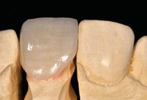
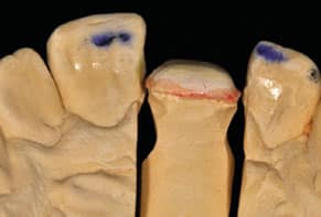
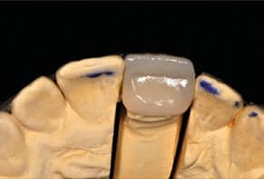
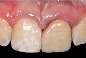
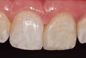
The final restoration/veneer was reviewed first on the working cast (Figure 6), and occlusion was checked carefully (Figure 7 and Figure 8). At the insertion appointment, marginal adaptation, restoration fit, interproximal contacts and occlusion were verified using a translucent try-in paste (Figure 9). Upon final approval by the patient, the internal bonding surface of the laminate veneers were etched with 10% hydrofluoric acid for 25 seconds and silanated. After the enamel was etched for 15 seconds, the veneer was luted with resin cement following the manufacturer’s recommendations for veneer cementation. All excess cement was removed from the margins and the restoration was cured with a curing light.24 Figure 10 shows the final cemented veneer at the follow-up appointment.
DISCUSSION
Treatment of severely dilacerated maxillary central incisors is a challenge, chiefly due to the difficult position and abnormality of the root. Therapy often involves surgical removal, with subsequent orthodontic treatment to either close the space or maintain space for further restorative treatment. A dilacerated tooth is more resistant to extrusion than a normal tooth and has a higher chance of apical resorption.15 While impacted maxillary central incisors with severe dilaceration can be treated with orthodontic therapy in some cases, restorative treatment may be necessary for an optimal esthetic outcome.14,15
Replacing a missing single tooth with implant-supported crown is a common, acceptable and reliable treatment,25 and the 94.5% success rate makes it an attractive option.26 In addition to a high success rate, this approach does not required preparation of adjacent teeth — and this is important in young patients with unrestored dentition and large pulps.27,28 However, the growth factor must be considered when placing implants in young patients. This is because placing implants in young adults whose growth continues may cause esthetic and functional challenges as they get older. Studies have shown that osseointegrated implants do not follow the eruptive path of adjacent natural dentition. This will cause progressive infraposition of implant-supported crowns and discrepancy of surrounding tissue.19–22 The skeletal maturation assessment did not confirm the complete growth of the patient presented in this clinical report, and, as a result, an implant-supported crown was not considered to be a treatment with a predictable outcome.29
A paper presenting three cases proposed direct composite restoration for maxillary central incisors with crown dilaceration.14 Although direct composite resin may provide an acceptable and cost-effective option, porcelain (i.e., ceramic) veneers have a higher survival rate and patient satisfaction.30 Thus, a glass ceramic with a full bonding cementation protocol was the treatment option chosen for this patient.
CONCLUSION
Paying close attention to occlusion and anterior guidance is essential when restoring maxillary central incisors with severe dilaceration. In this clinical scenario, occlusion in maximum intercuspation and anterior guidance were not effected by the treatment (as illustrated in Figure 7 and Figure 8).
A maxillary central incisor with a dilacerated crown compromises the patient’s esthetics and function, and could be challenging to treat. While many treatment options are available, using a conservative approach for properly selected patients — such as a bonded ceramic veneer — can restore function and esthetics with a predictable outcome.
KEY TAKEAWAYS
- Treatment of severely dilacerated maxillary central incisors is a challenge, chiefly due to the difficult position and abnormality of the root.
- Trauma is a common cause of root dilacerations, particularly in the maxillary anterior region, and various treatment approaches may be necessary — including orthodontic, surgical, endodontic and restorative therapies.8–13
- Paying close attention to occlusion and anterior guidance is essential when restoring maxillary central incisors with severe dilaceration.
- Using a conservative approach for properly selected patients — such as a bonded ceramic veneer — can restore esthetics and function with predictable outcomes.
REFERENCES
- The glossary of prosthodontic terms: Ninth edition. J Prosthet Dent. 2017;117(5S):e32–e105.
- Stewart DJ. Dilacerated unerupted maxillary central incisors. Br Dent J. 1978;145:229–233.
- Andreasen JO, Ravn JJ. Epidemiology of traumatic dental injuries to primary and permanent teeth in a Danish population sample. Int J Oral Surg. 1972;1:235–239.
- Andreasen JO, Sundström B, Ravn JJ. The effect of traumatic injuries to primary teeth on their permanent successor. I. A clinical and histologic study of 117 injured permanent teeth. Scand J Dent Res. 1971;79:219–283.
- Andreasen JO, Ravn JJ. The effect of traumatic injuries to primary teeth on their permanent successor. II. A clinical and radiographic follow-up study of 213 teeth. Scand J Dent Res. 1971;79:281–291.
- Andreasen JO. The influence of traumatic intrusion of primary teeth on their permanent successors. A radiographic and histological study in monkeys. Int J Oral Surg. 1976;5:207–219.
- Malcić A, Jukić S, Brzović V, Miletić I, Pelivan I, Anić I. Prevalence of root dilaceration in adult dental patients in Croatia. Oral Surg Oral Med Oral Pathol Oral Radiol Endod. 2006;102:104–109.
- Topouzelis N, Tsaousoglou P, Pisoka V, Zouloumis L. Dilaceration of maxillary central incisor: a literature review. Dent Traumatol. 2010;26:427–433.
- Singh H, Kapoor P, Sharma P, Dudeja P, Maurya RK, Thakkar S. Interdisciplinary management of an impacted dilacerated maxillary central incisor. Dent Press J Orthod. 2018;23:37–46.
- Bhikoo C, Xu J, Sun H, Jin C, Jiang H, Hu R. Factors affecting treatment duration of labial inversely impacted maxillary central incisors. Am J Orthod Dentofacial Orthop. 2018;153:708–715.
- Achary RC, Ravi GR. A novel approach of esthetic management and preserving vitality of dilacerated permanent maxillary lateral incisor. Int J Clin Pediatric Dent. 2016;9:152–155.
- Campbell CM, DiBiase A, Fleming PS. Concomitant dilaceration, transposition, and intraosseous migration: report of a patient treated with maxillary canine-central incisor substitution. Am J Orthod Dentofac Orthop. 2014;146:514–521.
- Sharma S, Grover S, Sharma V, Srivastava D, Mittal M. Endodontic and esthetic management of a dilacerated maxillary central incisor having two root canals using cone beam computed tomography as a diagnostic aid. Case Rep Dent. 2014:2014;861942.
- Mellara Tde S, Nelson-Filho P, Queiroz AM, Santamaria M, Silva RA, Silva LA. Crown dilaceration in permanent teeth after trauma to the primary predecessors: report of three cases. Braz Dent J. 2012;23:591–596.
- Pavlidis D, Daratsianos N, Jäger A. Treatment of an impacted dilacerated maxillary central incisor. Am J Orthod Dentofacial Orthop. 2011;139:378–387.
- Muthumani T, Rajasekaran M, Veerabahu M, Indra R. Interdisciplinary management of impacted maxillary central incisor with dilacerated crown. J Endod. 2011;37:269–271.
- Subramaniam P, Naidu P. Treatment of crown dilaceration: an interdisciplinary approach. Indian Soc Pedod Prev Dent. 2010;28:34–37.
- Chaugule V, Bhat C, Patil V, Shah K, Vihapure S. Endo-esthetic management of permanent maxillary lateral incisor with severe root dilaceration in a 12-year-old child. Int J Clin Pediatr Dent. 2008;1:32–38.
- Oesterle LJ, Cronin RJ Jr, Ranly DM. Maxillary implants and the growing patient. Int J Oral Maxillofac Implants. 1993;8:377–387.
- Cocchetto R, Canullo L, Celletti R. Infraposition of implant-retained maxillary incisor crown placed in an adult patient: case report. Int J Oral Maxillofac Implants. 2018;33:107–111.
- Mankani N, Chowdhary R, Patil BA, NagaraJ E, Madalli P. Osseointegrated dental implants in growing children: a literature review. J Oral Implantol. 2014;40:627–631.
- Cronin RJ Jr, Oesterle LJ. Implant use in growing patients. Treatment planning concerns. Dent Clin North Am. 1998;42:1–34.
- Fishman LS. Radiographic evaluation of skeletal maturation. A clinically oriented method based on hand-wrist films. Angle Orthod. 1982;52:88–112.
- Zandinejad A, Lin WS, Atarodi M, Abdel-Azim T, Metz MJ, Morton D. Digital workflow for virtually designing and milling ceramic lithium disilicate veneers: a clinical report. Oper Dent. 2015;40:241–246.
- Mayer TM, Hawley CE, Gunsolley JC, Feldman S. The single-tooth implant: a viable alternative for single-tooth replacement. J Periodontol. 2002;73:687–693.
- Jung RE, Pjetursson BE, Glauser R, Zembic A, Zwahlen M, Lang NP. A systematic review of the 5-year survival and complication rates of implant supported single crowns. Clin Oral Implants Res 2008;19:119−130.
- Kinzer GA, Kokich VO. Managing congenitally missing lateral incisors. Part III: single-tooth implants. J Esthet Restor Dent. 2005;17:202−210.
- Prado N, Malik O, Waring D. The management of the dilacerated impacted maxillary central incisor. Dent Update. 2016;43:618–630.
- Fishman LS. Maturational patterns and prediction during adolescence. Angle Orthod. 1987;57:178–193.
- Wakiaga J, Brunton P, Silikas N, Glenny AM. Direct versus indirect veneer restorations for intrinsic dental stains. J Esthet Restor Dent. 2006;18:111–113.
The authors have no commercial conflicts of interest to disclose.
From Decisions in Dentistry. June 2019;5(6):10,12,14,17.
