

Understanding the Clinician’s Role in Forensic Dentistry
With adequate training, dental teams can play an integral role in forensic odontology by identifying human remains, as well as possible signs of abuse or neglect.
With adequate training, dental teams can play an integral role in forensic odontology by identifying human remains, as well as possible signs of abuse or neglect
PURCHASE COURSE
This course was published in the July 2018 issue and expires July 2021. The author has no commercial conflicts of interest to disclose. This 2 credit hour self-study activity is electronically mediated.
EDUCATIONAL OBJECTIVES
After reading this course, the participant should be able to:
- Define forensic dentistry and its applications.
- Discuss the use of dental records for purposes of human identification.
- Explain how prints found on the lips can aid forensic odontology.
Forensic dentistry, or forensic odontology, focuses on the examination, evaluation, management and presentation of dental evidence in civil or criminal legal proceedings.1,2 It can be used to identify human remains in individual deaths or mass disaster events, note signs and symptoms of abuse or neglect, help to estimate approximate age, and assess a subject‘s oral cavity.3 While forensic dentistry is a specialized field, dentists in private practice play an important role by maintaining accurate dental records that may be used for identification in future cases.
Forensic dentistry is integral to the identification of individuals in murder investigations, abuse cases, possible criminal suspects and missing persons. Forensic dentists identify human remains through the comparison of antemortem and postmortem dental records. Forensic odontology can facilitate the identification of an individual after prolonged exposure to the environment, when advanced decomposition has occurred. As noted, it can also be used in identifying signs and symptoms of abuse or neglect, such as domestic, child and elderly abuse.
This field is also useful in identifying victims of mass fatalities due to accidents, acts of terror or natural disasters.4 For example, when records or fingerprints are not available, teeth can be used to identify an individual, because the dentition typically remains in some capacity, even when an individual is burned, decomposed or dismembered. Through an analysis of the dentition, existing restorations can be matched with dental records, age can be estimated, and anatomical appearance can be recreated.
ROLE OF DENTAL RECORDS
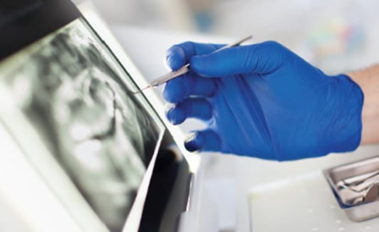
While dentists may not work directly at the scene of a traumatic event, each member of the dental team is important in ensuring records are valid and up to date, as this information may be necessary for identification. Dental personnel should be thorough and precise when documenting each examination or procedure. Records that may be used for identification purposes include medical history, dental history, dental examination info, radiographs, intra- and extraoral photographs, study casts, treatment plans and treatment entries (Figure 1).
Accurate records must be maintained at each visit. Appropriate dental documentation should include treatment, restorative care, dental anatomy and periodontal status. Dental records should be legible, well maintained and accessible when requested by the appropriate authorities.
TEETH AS IDENTIFIERS
The oral cavity provides myriad ways of aiding the forensic identification process. Bone examination can be used for reconstruction purposes. In addition to the facial bones, cone beam computed tomography, radiographs and photographs can be used to reconstruct an individual’s face. The presence of implants and joint replacements can also help identify an individual; similarly, salivary DNA analysis may also be used.5
The presence or absence of particular teeth and the position of various teeth can aid identification. Exact and correct documentation is paramount, as this information could be used for comparative purposes when needed to identify an individual.
Handheld X-ray machines can be used at a site where a body is found or within a morgue to produce intraoral radiographs. These machines are lightweight and designed to accommodate possible inadvertent movement that may occur when using a handheld device.4 Radiographic imaging collected through this process on the postmortem body can be compared to antemortem radiographs for similarities or differences. Imaging collected via a radiographic sensor connected to a laptop or desktop computer can be immediately viewed and compared to antemortem images.6
POSTMORTEM DENTAL EXAM
Problems that may arise when identifying an individual through dental records include:
- Clinical records that may not be documented properly or updated at subsequent appointments.
- Variances in the information present, as there is no standardization with charting systems. An office could be using universal, Palmer, or the Fédération Dentaire Internationale (FDI) system to identify the dentition. It is imperative to know which identification system the office is using.
- Radiographs that could be mounted incorrectly.
- Family and friends who are not aware of the dental office used by the individual in question, thus complicating efforts to compare antemortem and postmortem dental records.
The postmortem dental examination is conducted by the authority and under the direction of the coroner/medical examiner or the designee, typically a forensic odontologist.7 Examinations are usually conducted in a morgue setting or temporary station set up at the scene of a mass disaster. Once the body is ready for evaluation, computer equipment can be used while information is collected for the comparison of postmortem and antemortem data. The tools used by the examiner to analyze and collect data include items typical of routine dental examinations, such as intraoral cameras, handheld radiography units, and computers with programs for item analysis. Remains are charted using a description that is similar to an intraoral examination.
Any identifying factors from an oral exam should be carefully documented. Once the information is collected, a database known as WinID contains antemortem information of missing persons that can be used for forensic comparison purposes. Both postmortem and antemortem documentation must be thorough and accurate.6 There are four possible results of a forensic comparison evaluation: positive identification, presumptive (probable) identification, insufficient identification evidence, and exclusion of identification evidence.
Positive identification occurs when no major antemortem and postmortem differences are seen in the evidence.
Presumptive (probable) identification is when there are similarities between antemortem and postmortem items, yet there is the possibility that some information is absent. A total positive identification may not occur when probable identification is the result.
Insufficient identification evidence is when it is difficult to arrive at a conclusion when antemortem and postmortem items are compared because there is not enough evidence to make such an identification.
Exclusion of identification evidence reveals explainable or unexplainable differences that occur in antemortem or postmortem items, which lead to inconsistencies.8
Professionals who examine individuals in the postmortem state should always follow federal Occupational Safety and Health Administration protocols and accepted infection control standards. Occlusal images of both dental arches, full face and head views, and unusual restorative features should be photographed. The examiner should also conduct a comprehensive evaluation of all antemortem records. Once all information is obtained, a comparison analysis should be conducted to formulate a conclusion.9
METHODS AND TECHNIQUES

over a lifetime, making cheiloscopy a useful forensic identification tool.NADYAPHOTO/ISTOCK/GETTY IMAGES PLUS
Cheiloscopy is a forensic identification technique that can be used to verify identify through prints found on the lips, as these groove patterns are unique for each person and rarely change throughout the life of an individual. These imprints may be found on a glass after drinking, on cigarette butts, and a window if pressed upon with the lips (Figure 2). Suzuki and Tsuchihashi10 classified lip prints into six categories for analysis, based on the various appearances of grooves seen on the lips:
- Type I states there are clear-cut grooves running vertically across the lip.
- Type I’ indicates the grooves are straight, but disappear halfway instead of covering the entire length of the lip.
- Type II shows grooves that fork in their course.
- Type III suggests the grooves intersect.
- Type IV promotes the reticulation of grooves.
- Type V provides no indication of anything seen in types I to IV, thus, they cannot be differentiated morphologically.
These images can be lifted for analysis and comparison through the use of aluminum powder and magnetic bonds.11
Another identification approach is rugoscopy. This technique uses the shape, length, number and direction of the rugae found on the palatal surface of the mouth to help identify an individual. The advantage to using rugae is that they are usually protected inside the oral cavity.12
As noted, teeth have a strong resistance to decomposition and damage when compared with other body tissues. Because of this resistance, teeth can contain DNA when it is not available elsewhere. Pulp is the ideal area of the tooth from which to obtain a DNA sample. Dentin and cementum can also be used to collect DNA in cases in which the pulp is not present, such as when root canal therapy has been performed.13
ADDITIONAL APPLICATIONS
Forensic dentistry is particularly important in the presence of mass fatalities. Various response teams are summoned when a mass disaster occurs, including the Disaster Mortuary Operational Response Team (perhaps more commonly know by the acronym DMORT). This group may include medical examiners, coroners, dentists, dental hygienists and dental assistants.14
As previously noted, forensic dentistry is also useful in determining whether abuse or neglect has occurred. The current condition of the individual can be compared with previous records before the abuse occurred. Members of the dental team can evaluate the oral cavity to see if the anatomy has changed in ways that may be indicative of abuse. For example, bruises/burns on the lips or within the oral cavity, fractured teeth and damaged tissue, — as well as bite marks — can all be indicators of abuse.
Bite marks can be evaluated to identify the perpetrator of the bite. While most often found on victims, bite marks may also be found on the perpetrator if the victim bit in self-defense.15 Photos of the area in question, impressions, a sample bite and study casts all should be collected to use for comparative purposes. The evaluation of bite marks was considered a standard in forensic dentistry, yet recent cases have led to questions about its validity. In several high-profile cases in recent years, bite mark evidence has been proven invalid when compared with subsequent DNA analysis.16 Based on DNA exoneration, studies of wrongful convictions have found the forensic sciences to be second only to eyewitness errors as a source of false or misleading evidence.17 As cases are further examined, the forensic odontologist should be thorough when using this approach due to the ramifications if an incorrect conclusion is reached.
Age assessment can be performed through attrition, periodontal attachment levels, root resorption, secondary dentin, and cementum apposition. Estimation of the age of human remains can assist in determining identification. Measurements of root transparency, which require sectioning of a tooth, or measuring and counting certain internal anatomical landmarks within a tooth, are some of the characteristics evaluated during forensic age assessment.16
Another approach that can be used in forensic dentistry is facial reconstruction. In some instances, the only portion of the body remaining is the skeleton, including the skull. The skull is scanned with a laser camera and software is used to create a three-dimensional (3D) virtual facial image.18 In addition, 3D printing can be used to create models of anatomical structures representing bone fractures, vessels, ruptured organs and bite marks. These replicas can be presented to discuss forensic findings and victim identification in the courtroom.19 A structure is scanned, software interprets the image, and the information is relayed to a 3D printer. The printed copy can be handled by multiple individuals without harming or damaging the original item. Images and replicas can be constructed of select features, such as bite marks, before changes begin to take place with the initiation of healing.3 Besides helping to identify fracture patterns or facial features, 3D printing has also been used in the reconstruction and identification of weapons used against victims.20
CONCLUSION
Forensic dentistry is key to helping identify individuals who may otherwise remain unidentified. The dental team plays a pivotal role in the identification process by maintaining accurate and precise antemortem documentation of oral examinations and treatment. Fellowship programs, continuing education courses, and workshops are all avenues where oral health professionals can learn more about opportunities in forensic odontology.
REFERENCES
- Krishan K, Kanchan T, Garg A. Dental evidence in forensic identification — an overview, methodology and present status. Open Dent J. 2015;9:250–256.
- Balachander N, Aravindha Babu N, Jimson S, Priyadharsini C, Masthan K. Evolution of forensic odontology: an overview. J Pharm Bioallied Sci. 2015;7:S176–S180.
- Khanna S, Dhaimade P. Exploring the 3rd dimension: Application of 3D printing in forensic odontology. J Forensic Sci Criminal Inves. 2017;3:1–3.
- Hinchiffe J. Forensic odontology, part 2. Major disasters. Br Dent J. 2011;210:269–274.
- Verma A, Kumar S, Rathore S, Pandey A. Role of dental expert in forensic odontology. Natl J Maxillofac Surg. 2014;5:2–5.
- Shanbhag V. Significance of dental records in personal identification in forensic sciences. J Forensic Sci Med. 2016;2:39–43.
- American Board of Forensic Odontology Diplomates Reference Manual. Available at: http://abfo.org/wp-content/uploads/2012/08/ABFO-Reference-Manual-April-2017-v7.pdf. Accessed May 21, 2018.
- Kenney J, Standish S, Souviron R, Vale G, McGivney J, Jones G. Body identification guidelines. J Am Dent Assoc. 1994;125:1244–1254.
- Pretty I, Sweet D. A look at forensic dentistry-part 1: The role of teeth in the determination of human identity. Br Dent J. 2001;190:359–366.
- Suzuki K, Tsuchihashi Y. Personal identification by means of lip prints. J Forensic Med. 1970;17:52–57
- Castelló A, Alvarez-Seguí M, Verdú F. Luminous lip-prints as criminal evidence. Forensic Sci Int. 2005;155:185–187.
- Shukla D, Chowdhry A, Bablani D, Jain P, Thapar R. Establishing the reliability of palatal rugae pattern in individual identification (following orthodontic treatment). J Forensic Odontostomatol. 2011;29:20–29.
- Ata-Ali J, Ata-Ali F. Forensic dentistry in human identification. J Clin Exp Dent. 2014;6:162–167.
- U.S. Department of Health and Human Services. Disaster Mortuary Operational Response Teams. Available at: phe.gov/Preparedness/responders/ndms/ndms-teams/Pages/dmort.aspx. Accessed May 21, 2018.
- Pallam NK, Boaz K, Natrajan S, Raj M, Manaktala N, Lewis AJ. Computer-based method of bite mark analysis: a benchmark in forensic dentistry? J Forensic Dent Sci. 2016;8:32–39.
- Metcalf R, Klim-Lemann J. Overview of forensic odontology. J Calif Dent Assoc. 2015;7:295–301.
- Oxford University Press USA. It’s time to stop using bite marks in forensics, experts argue. Available at: sciencedaily.com/releases/2016/12/161201120128.htm. Accessed May 21, 2018.
- Jeddy N, Ravi S, Radhika T. Current trends in forensic odontology. J Forensic Dent Sci. 2017;9:115–119.
- Newcomb TL, Bruhn AM, Giles B, Garcia NH, Diawara N. Testing a novel 3D printed radiographic imaging device for use in forensic odontology. J Forensic Sci. 2017;62:223–228.
- Woźniak K, Rzepecka-Woźniak E, Moskała A, Pohl J, Latacz K, Dybała B. Weapon identification using antemortem computed tomography with virtual 3D and rapid prototype modeling — a report in a case of blunt force head injury. Forensic Sci Int. 2012;222:e29–e32.
Featured image by PAULFLEET/ISTOCK/GETTY IMAGES PLUS
From Decisions in Dentistry. July 2018;4(7):38–41.

