

Multifactorial Periodontal Risk Assessment
Successful periodontal treatment depends on a clinician’s ability to identify, monitor and manage complex risk factors.
Successful periodontal treatment depends on a clinician’s ability to identify, monitor and manage complex risk factors
This course was published in the June 2016 issue and expires 06/30/19. The authors have no commercial conflicts of interest to disclose. This 2 credit hour self-study activity is electronically mediated.
OBJECTIVES
After reading this course, the participant should be able to:
- Describe the risk factors for periodontal disease.
- Explain the mechanisms that mediate risk factors in periodontal disease.
- Identify risk factors for periodontal disease in dental patients.
Periodontal disease is not universally distributed among all adult population. Analysis of the 2009–2010 National Health and Nutrition Examination Survey (NHANES) shows that 47% of U.S. adults age 30 and older had periodontitis, including 8.7% with mild disease, 30% with moderate periodontitis, and 8.5% with severe disease.1 An individual’s susceptibility to periodontal infection and/or the degree of host response to this infection account for these differences.
The recognition that differences in susceptibility are responsible for differences in prevalence and severity led to the study of factors associated with periodontal disease, independent of the effects of confounding variables. Independent risk factors include systemic diseases and conditions — such as diabetes mellitus, obesity, metabolic syndrome and osteoporosis — and lifestyle factors, including smoking and alcohol use. Since most risk factors are modifiable and are found in a large number of patients with periodontal disease, identifying and managing risk has become integral to therapy.
A risk factor is defined as a characteristic, aspect of behavior, or an environmental exposure that is associated with periodontitis (although the etiology remains subgingival microbial infection). Risk factors either increase the host’s susceptibility to infection or the inflammatory response to such infection. Modifiable risk factors are usually systemic, environmental or behavioral. Nonmodifiable risk factors, also known as determinants, are intrinsic to the individual — such as age and gender. Similar to successful management of chronic multifactorial diseases, identifying and managing risk factors in patients with periodontitis is essential for successful outcomes and long-term disease control.
MODIFIABLE RISK FACTORS
ENVIRONMENTAL FACTORS
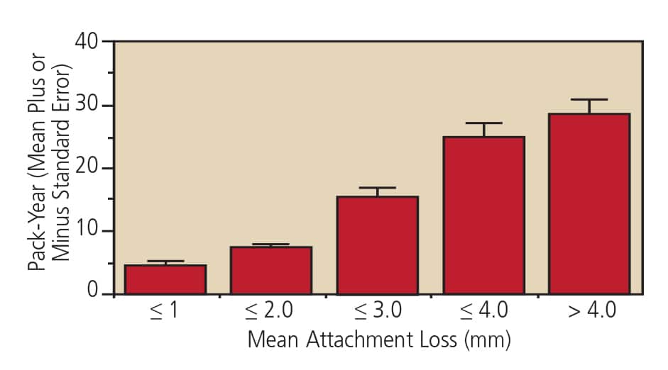
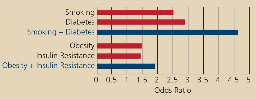
Smoking is the most important environmental — and modifiable — risk factor for periodontal disease. Studies that examined the effect of smoking on periodontal disease provide a clear association as follows: current and former smokers exhibit different risk profiles; in addition, the negative effect of smoking on periodontal health is cumulative and proportional to the amount of smoking. On average (and independent of the effect of age, gender and amount of dental plaque), smokers are at 2.5 times greater risk of developing severe periodontal disease than nonsmokers (never-smokers). The odds for periodontitis for current smokers are higher than for former smokers, 3.97 versus 1.68 respectively. The odds for developing periodontal disease range from 2.0 to 7.0, depending on number of cigarettes smoked per day and years of smoking).2 Furthermore, the severity of clinical attachment loss and alveolar bone loss is proportional to the lifelong history of smoking (Figure 1). Smoking presents a greater risk for progression of periodontitis than baseline level of disease. In a two-year longitudinal study, the odds for disease progression were 3.75 for smokers, compared to 2.29 for baseline clinical attachment level of >6mm.3
Cigarette smoking increases the risk for colonization with Porphyromonas gingivalis, Tannerella forsythia and Aggregatibacter actinomycetemcomitans.4 Current smokers have 2.3 greater odds to harbor these pathogens compared to former and never-smokers with comparable disease severity.4 Current smokers with periodontitis were more likely to be colonized with specific periodontal pathogens, including Campylobacter rectus5 and have 4.6 odds of harboring Treponema denticola in periodontal pockets.6 Major differences in the prevalence (i.e., % of sites colonized) of subgingival pathogens were seen in current smokers compared to former and never-smokers.7 The greater colonization in current smokers appears to be due to greater colonization at pocket depths <4 mm.
Current smokers showed also greater colonization by periodontal pathogens and exogenous bacteria, such Escherichia coli, Candida albicans, Aspergillus fumigatus and Staphylococcus aureus.8 Collectively, the data suggest that cigarette smoking adversely affects the host response to infection. P. gingivalis genotype fimA IV was associated with disease severity in current smokers.9 The fimA IV genotype is considered pro-inflammatory and aggressive due to the presence of capsule and fimbriae, which provides an advantage in terms of invasion, survival within the host, and resistance from clearance — all elements related to the chronicity and severity of periodontal infection in smokers.
Treatment studies report unpredictable results in removing periodontal pathogens in current smokers. Scaling and root planing (S/RP) is less effective at eliminating periodontal pathogens in current smokers compared to former smokers.10 A review of treatment studies concluded that, compared to nonsmokers, smokers consistently exhibited a poorer response to both nonsurgical (S/RP plus locally delivered antibiotics) and surgical therapy. This reduced treatment outcome was also seen at class I and II furcation sites.11 Smokers also have less success with open-flap debridement, osseous resection, soft tissue and bone grafting, and regenerative procedures. The response to periodontal therapy of former smokers suggests that quitting smoking is beneficial to treatment outcomes. Therefore, a smoking status assessment should be included in periodontal diagnosis, prognosis and treatment planning. Smoking cessation should be considered as part of periodontal treatment and should be encouraged for all current smokers.
SYSTEMIC FACTORS
Diabetes is the most significant systemic — and modifiable — risk factor for periodontal disease. Compared with other patient groups, periodontal disease is more common in individuals with diabetes — for example, young adults with diabetes are twice as likely to develop periodontal disease as those without diabetes. Individuals age 45 and older with poorly controlled (A1c >9) diabetes are 2.9 times more likely than those without diabetes to have severe periodontitis. The risk increases to 4.6 among current smokers with poorly controlled diabetes.12 Once periodontal disease is established in a diabetic host, the chronic nature of infection/inflammation constitutes a risk for difficulty in glucose control and diabetes severity. Thus, the relationship between diabetes and periodontal disease is two-way: diabetes increases the risk for periodontitis, while periodontal infection and inflammation make diabetes more difficult to control.13
Moderate to severe periodontitis is associated with an increased risk for macroalbuminuria, end-stage renal disease, calcification of atherosclerotic plaques, carotid intima-medial thickness, and even cardio-renal mortality.14 There is a direct and dose-dependent relationship between periodontitis severity and diabetes complications in both type 1 and type 2 diabetes, with emerging evidence that periodontitis may predispose individuals to diabetes.15–17
Guidelines for periodontal care and managing oral health have been proposed to physicians, dental practitioners and patients affected by, or at risk of, diabetes.18 These principles state that patients with diabetes should be educated that their risk of developing periodontal disease is high, and that if they are diagnosed with a periodontal condition, glycemic control may be difficult and poses a significant risk for complications. Periodontal examinations should be performed on all patients with diabetes. In fact, annual periodontal examinations are recommended, even in the absence of periodontitis.18
OBESITY AND METABOLIC SYNDROME
The most important risk factor for type 2 diabetes is obesity. A systematic review and meta-analysis of the literature concluded that obesity — measured as increased body mass index (BMI) — is associated with increased severity of periodontal disease.19 Insulin resistance mediates the increased risk for type 2 diabetes and periodontal disease in obese individuals.20 Accordingly, waist circumference — a measure of central obesity/abdominal fat more closely related to insulin resistance than whole body BMI — is more closely associated with periodontal disease than raw BMI score. The increased risk for periodontal disease associated with obesity is proportional to the degree of obesity, with the risk increasing for every additional centimeter of waist circumference or unit of BMI score.
Metabolic syndrome also significantly increases the risk of developing cardiovascular disease and diabetes. A combination of factors, including abdominal obesity, insulin resistance, abnormal lipids and hypertension, contributes to this risk. The association between periodontitis and metabolic syndrome has been reported, though the possible confounding effect of diabetes risk has not been adequately addressed in most published reports. D’Aiuto et al examined data from the NHANES III study and found an association between metabolic syndrome and severe periodontitis only in individuals older than 45.21
OSTEOPOROSIS AND OSTEOPENIA
Osteopenia is a systemic condition in which bone mineral density (BMD) falls below a predefined level considered adequate for age and gender. Contrarily, osteoporosis is a disease in which BMD is below the levels required for mechanical support. Solid evidence supports the role of osteopenia and osteoporosis as risk factors for tooth loss in both dentate and edentulous individuals.22,23 Krall et al found that for each 1%-per-year decrease in whole body BMD, the risk for tooth loss more than quadrupled in postmenopausal women.23 Although the evidence supporting osteoporosis and osteopenia as risk factors for periodontal disease is not as clear, it nevertheless points to an association between osteopenia, osteoporosis and periodontal disease.24,25 This association is mediated by estrogen, calcium and vitamin D. Information on skeletal BMD, hormonal and vitamin supplementation status should be ascertained as part of a complete medical history of postmenopausal periodontal patients.
DIETARY RISK FACTORS
Analyses of large population-based datasets reveals that reduced calcium intake and low serum calcium levels are associated with increased risk for periodontal disease.26 Low dietary calcium intake has been associated with increased attachment loss in a dose-dependent manner in a representative sample of the U.S. population.27 Low dietary intake of vitamins C and D are also associated with increased risk for periodontal disease.27,28 Low levels of vitamin D intake result in decreased serum levels of calcium, which, in turn, stimulate the parathyroid gland to produce parathyroid hormone — leading to increased osteoclastogenesis.29 Assessing dietary intake of calcium and vitamins C and D should be included in every periodontal exam.
ALCOHOL CONSUMPTION
Alcohol consumption is associated with a moderate risk of periodontal disease (30%), as measured by gingival recession and clinical attachment loss.30,31 Periodontal disease risk is associated with the amount of alcohol consumed, not the type of alcohol. Increased risk of periodontal disease is seen in individuals who drink more than five alcoholic beverages per week, compared to those who drink less or not at all. The risk increases to 40% for individuals who consume more than 10 alcoholic drinks per week. The increased risk is independent of the behavioral aspect associated with alcohol — such as poor oral hygiene — and likely mediated by the multiple effects alcohol has on susceptibility to infection, inflammation and immune response.30,31
PSYCHOLOGICAL FACTORS
Psychological stress, distress and depression have been reported to increase the risk for periodontal disease.32 A large population-based study reported that individuals experiencing high financial stress exhibited increased severity of periodontal disease compared to those with less financial stress.32 A systematic review of available evidence reports that cross sectional and case control studies showed a positive correlation between stress and periodontal disease. Only two of the studies reviewed were prospective clinical studies, and both showed a relationship between stress and periodontal disease.33 Very few studies have examined coping mechanisms associated with stress and distress. Coping behaviors modified the negative effects of stress on periodontal tissues; that is, individuals with high levels of problem-focused coping where at less risk of periodontal disease than individuals with high stress and emotion-focused coping (a form of inadequate coping).32
Stress, distress and depression may be associated with periodontal disease through behavioral and physiologic mechanisms. Behavioral mechanisms include poor oral hygiene, unhealthy diet, and other behaviors not conducive to oral health. Physiologic mechanisms are mediated by cortisol, a hormone secreted during stress that affects inflammations. Consequently, assessing stress, distress, depression and coping behaviors should be included in periodontal practice.
NONMODIFIABLE RISK FACTORS
GENETIC FACTORS
There is evidence suggesting the role of genetic factors in aggressive periodontitis. The genetic markers responsible for the increased risk include polymorphisms in the IL-1B gene, which is responsible for up to threefold increases in IL-1 production.34 Polymorphisms at the IL-1A and IL-1B genes are associated with chronic adult periodontitis only in nonsmokers. In smokers, severe periodontal disease was not associated with any of the examined candidate polymorphisms. A study of Italian adults with periodontitis and healthy controls reported polymorphisms of IL 6 and IL1 genes associated with periodontitis, while no there was no association with IL1A or ILB genotype.35 Although genetic factors, such as specific gene polymorphisms, are suspected to have an association with chronic adult periodontitis, there is yet no clear evidence for this in the general population.
BACKGROUND FACTORS
Age and gender are considered background (or nonmodifiable) risk factors. Studies of the natural history of periodontal disease show that severity of periodontal destruction increases with age.36,37 In the Erie County study (a sample representative of the Western New York population) clinical attachment loss2 and alveolar bone loss severity38 were directly proportional to increases in age. Data from NHANES 2009 and 2010 indicate that 64% of adults age 65 years and older had moderate or severe periodontitis, compared to 47% of subjects ages 30 to 64.1 Periodontal disease is also more prevalent and more severe in men than in women of comparable age.1,2,38
ASSESSING AND MODIFYING RISK FACTORS
The concept of risk assessment has been incorporated into the American Academy of Periodontology guidelines for managing patients with periodontal disease (Figure 2). In fact, risk assessment should be part of every comprehensive and periodic dental and periodontal examination. Managing risk factors and reducing risk is part of contemporary periodontal care.
Meta-analysis of studies of periodontal treatment in patients with diabetes report reductions in levels of HbA1c of -0.38%, -0.66% when restricted to type 2 diabetic patients, and -0.71% if antibiotics were given.39 A subsequent meta-analysis of treatment studies demonstrated a weighted mean reduction in HbA1c levels of -0.40% for at least three months after periodontal treatment.40 This evidence underscore the importance of periodontal treatment in diabetic patients.
Other strategies to reduce risk for periodontal disease include emphasizing the importance of weight management, and promoting healthy lifestyles among obese patients. The importance of adequate diet and nutrient intake, and strategies for reducing stress and improving coping mechanisms are also part of risk factor modification. Ultimately, identifying and managing risk factors will help ensure optimal oral health and treatment outcomes in patients with — or at risk of — periodontal disease.
References
- Eke PI, Dye BA, Wei L, Thornton-Evans GO, Genco RJ et al. Prevalence of periodontitis in adults in the United States: 2009 and 1999–2010. J Dent. Res. 2012;91(10):914–920.
- Grossi SG, Zambon JJ, Ho AW, Koch, et al. Assessment of risk for periodontal disease. I. Risk indicators for attachment loss. J Periodontol. 1994;65:260–267.
- Ogawa H, Yoshijara A, Hirotomi T, Ando Y, Miyazaki H. Risk factors for periodontal disease progression among elderly people. J Clin Periodontol. 2002;29(7):592–597.
- Zambon JJ, Grossi SG, Machtei EE, Ho AW, Dunford R, Genco RJ. Cigarette smoking increases the risk for subgingival infection with periodontal pathogens. J Periodontol. 1996;67:1050–1054.
- Kubota M, Tanno-Nakanishi M, Yamada S, Okuda K, Ishihara K. Effect of smoking on subgingival microflora of patients with periodontitis in Japan. BMC Oral Health. 2011;11:1.
- Umeda M, Chen C, Bakker I, Contreras A, Morrison JL, Slots J: Risk indicators for harboring periodontal pathogens. J Periodontol. 1998;69(10):1111–1118.
- Haffajee AD, Socransky SS: Relationship of cigarette smoking to the subgingival microbiota. J Clin Periodontol. 2001;28(5):377–388.
- Kamma JJ, Nakou M, Baehni PC: Clinical and microbiological characteristics of smokers with early onset periodontitis. J Periodontal Res. 1999;34(1):25–33.
- Texeira SR, Mattarazo F, Feres M, Figuereido LC, de Faveri M, Simionato MR, Mayer MP. Quarification of Porphyromonas gingivalis and fimA genotypes in smoker chronic periodontitis. J Clin Periodontol. 2009;36(6):482–487.
- Grossi SG, Zambon JJ, Machtei EE, et al. Effect of smoking and smoking cessation on healing after mechanical periodontal therapy. J Am Dent Assoc. 1997;128:599–607.
- Heasman L, Stacey F, Preshaw PM, McCracken GI, Hepburn S, Heasman PA. The effect of smoking on periodontal treatment response: a review of clinical evidence. J Clin Periodontol. 2006;33(4):241–253.
- National Diabetes Fact Sheet, 2011: Diabetes Public Health Resource. U.S. Centers for Disease Control and Prevention. Available at: http://www.cdc .gov/ diabetes/ pubs/ pdf/ ndfs_ 2011.pdf. Accessed May 17, 2016.
- Grossi SG, Genco RJ. Periodontal disease and diabetes mellitus; a two-way relationship. Ann Periodontol. 1998;3:51–61.
- Sareni A, Nelson RG, Tulloch-Reid M, et al. Periodontal disease and mortality in type 2 diabetes. Diabetes Care. 2005;28:27–32.
- Thortensson H, Kuylenstierna J, Hugoson A. Medical status and complications in relation to periodontal disease experience in insulin-dependent diabetics. J Clin Periodontol. 1996;23:194–202.
- Shultis WA, Weil EJ, Looker HC, et al. Effect of periodontitis on overt nephropathy and end-stage renal disease in type 2 diabetes. Diabetes Care. 2007;30:306–311.
- Saito T, Shimazaki Y, Kiyohara Y, et al. The severity of periodontal disease is associated with the development of glucose intolerance in nondiabetics: the Hiyasama study. J Dent Res. 2004;83:485–490.
- Chapple IL, Genco R, working group 2 of the joint EFP/AAP workshop. Diabetes and periodontal diseases: consensus report of the Joint EFP/AAP Workshop on Periodontitis and Systemic Diseases. J Periodontol. 2013;84(Suppl 4):S106–S112.
- Chaffee BW, Weston SJ. Association between chronic periodontal disease and obesity: a systematic review and meta-analysis. J Periodontol. 2010;81:1708–1724.
- Genco RJ, Grossi SG, Ho AW, Nishimura F, Murayama Y. A proposed model linking inflammation to obesity, diabetes and periodontal infections. J Periodontol. 2005;76(Suppl 11):2075–2084.
- D’Aiuto F, Sabbah W, Netuveli G, Donos N, et al. Association of the metabolic syndrome with severe periodontitis in a large U.S. population-based survey. J Clin Endocrinol Metab. 2008;93:3989–3994.
- Krall EA, Dawson-Hughes B, Papas A, Garcia RI. Tooth loss and skeletal bone density in healthy postmenopausal women. Osteoporos Int. 1994;4:104–109.
- Krall EA, Garcia RI, Dawson-Hughes B. Increased risk for tooth loss is related to bone loss at the whole body, hip and spine. Calcif Tissue Int. 1996;59:433–437.
- Tezal M, Wactawski-Wende J, Grossi SG, Ho AW, Dunford R, Genco RJ. The relationship between bone mineral density and periodontitis in postmenopausal women. J Periodontol. 2000;71:1492–1498.
- Wactawski-Wende J, Hausmann E, Hovey K, Trevisan M, Grossi SG, Genco RJ. The association between osteoporosis and alveolar crestal height in postmenopausal women. J Periodontol. 2005;76(Suppl 11):2116–2124.
- Nishida M, Grossi SG, Dunford RG, Ho AW, Trevisan M, Genco RJ. Calcium and the risk for periodontal disease. J Periodontol. 2000;71:1057–1066.
- Nishida M, Grossi SG, Dunford RG, Ho AW, Trevisan M, Genco RJ. Dietary vitamin C and the risk for periodontal disease. J Periodontol. 2000;71:1215–1223.
- Hildebolt CF. Effect of vitamin D and calcium on periodontitis. J Periodontol. 2005;76:1576–1587.
- Millen AE, Hovey KM, Lamonte MJ. Plasma 25-hydroxyvitamin D concentrations and periodontal disease in postmenopausal women. J Periodontol. 2013;84:1243–1256.
- Tezal M, Grossi SG, Ho AW, Genco RJ. The effect of alcohol consumption on periodontal disease. J Periodontol. 2001;72:183–189.
- Tezal M, Grossi SG, Ho AW, Genco RJ. Alcohol consumption and periodontal disease: The Third National Health and Nutrition Examination Survey. J Clin Periodontol. 2004;31:484–488.
- Genco RJ, Ho AW, Grossi SG, Dunford RG, Tedesco LA. Relationship of stress, distress and inadequate coping behaviors to periodontal disease. J Periodontol. 1999;70:711–723.
- Halawany HS, Abraham NB, Jacob V, Al Amri MD, Patil S, Anil S. Is psychological stress a possible risk factor for periodontal disease? A systematic review. J Psychiatry. 2015;18:217.
- Kornman KS, Crane A, Wang HY, et al. The interleukin-1 genotype as a severity factor in adult periodontal disease. J Clin Periodontol. 1997;24:72–77.
- Scapoli L, Girardi A, Palmieri A, Carinci F, Testori T, Zuffetti F et al. IL6 and IL10 are genetic susceptibility factors of periodontal disease. Dent Res J. 2012;(2):S197–S201.
- Ismail AI, Morrison E, Burt BA, et al. Natural history of periodontal disease in adults: findings from the Tecumseh Periodontal Disease Study, 1959–1987. J Dent Res. 1990;69:430–435.
- Neely A, Holford TR, Loe H, Anerud A, Boysen H. The natural history of periodontal disease in man. Risk factors for progression of attachment loss in individuals receiving no oral health care. J Periodontol. 2001;72:1006–1015.
- Grossi SG, Genco RJ, Machtei EE, et al. Assessment of risk for periodontal disease II. Risk indicators for alveolar bone loss. J Periodontol. 1995;66:23–29.
- Janket SJ, Wightman A, Baird AE, Van Dyke TE, Jones JA. Does periodontal treatment improve glycemic control in diabetic patients? A meta-analysis of intervention studies. J Dent Res. 2005;84(12):1154–1159.
- Teeuw WJ, Gerdes VE, Loos BG. Effect of periodontal treatment on glycemic control of diabetic patients: a systematic review and meta-analysis. Diabetes Care. 2010;33(2):421–427.

