
Interpreting Occlusion
The role of assessment and patient education in preventing occlusal problems.
The role of assessment and patient education in preventing occlusal problems
Occlusion can seem like a complex and confusing subject. Temporomandibular disorders (TMDs), centric relation, maximum intercuspation, working and nonworking interferences, canine guidance, disk displacement and parafunctional habits all affect the dentition, and related disturbances can interfere with patients’ masticatory systems. Dental professionals should have an understanding of tooth movement so they can effectively advise their patients and plan appropriate treatment.
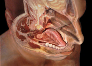
The masticatory system is composed of the structures required for adequate chewing, including the jaw and its related muscles, teeth, temporomandibular joints, tongue, lips, cheeks and mucous membranes (Figure 1). The teeth are just one part of the masticatory system, and if they are not in total equilibrium with the rest of the system, problems are likely to occur.1 Dental professionals have the opportunity to intercept failures in the masticatory system by observing certain signs and symptoms during routine dental appointments. With a basic understanding of relevant concepts and principles, clinicians can alert patients to possible problems.
TEMPOROMANDIBULAR DISORDERS
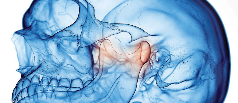
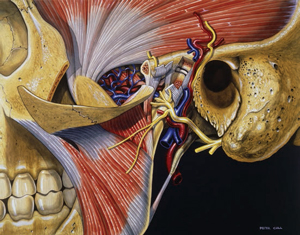
TMDs affect the masticatory muscles and the temporomandibular joint (TMJ), resulting in pain and/or dysfunction.2 TMJ and TMD are often used interchangeably when, in fact, TMD refers to the dysfunction and TMJ refers to the joint’s anatomical structure (Figure 2).
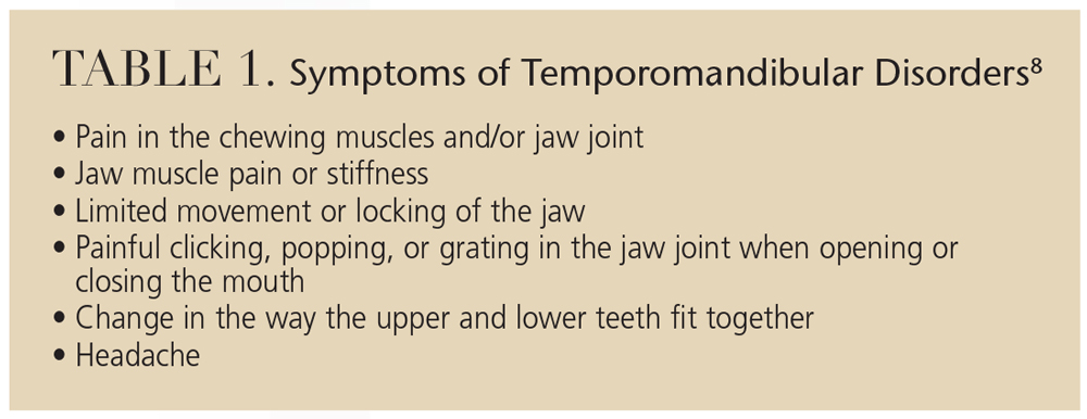
TMDs are the most common causes of nontooth-related chronic orofacial pain, with a prevalence between 5% and 12%.3–7 TMDs are more common among young people, particularly women.4–7 Risk is increased among women who take an estrogen supplement or use oral contraceptives.4–7 Table 1 provides a list of symptoms.8
CENTRIC RELATION
ANATOMICAL TRAVELOGUE/SCIENCE SOURCE
Centric relation (CR) is defined as “the relationship of the mandible to the maxilla when the properly aligned condyle-disk assemblies are in the most superior position against the eminentiae, irrespective of vertical dimension or tooth position.”1 In other words, CR is the position of the mandible in relation to the maxilla when everything is in the correct physiologic position. Tooth position and occlusion do not determine CR, as it is strictly a joint position.
MAXIMUM INTERCUSPATION
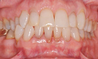
Formerly known as centric occlusion, maximum intercuspation (MI) occurs when the maxillary and mandibular teeth are fully interposed, that is, the position of maximum closure of the mouth (Figure 3). CR and MI are often used interchangeably, when, in reality, they are distinct. While CR is an optimal joint position, MI refers to a relationship between teeth. In order to gain occlusal harmony, the aim is to make MI coincide with CR, so that when the teeth are fully interposed, the condyles are properly seated in the joint space with the disk properly in place.
WORKING AND NONWORKING INTERFERENCES
When CR and MI do not coincide, working and nonworking interferences can occur that cause the mandible to deviate from its normal physiologic position. A working interference occurs when the teeth contact prematurely on the side where the active chewing or biting is taking place. In other words, if the teeth are brought together and the mandible shifts to one side, the working interference will be on the side of the mouth that the mandible is sliding toward. A nonworking interference occurs on the opposite side from where the mandible is sliding.
CANINE GUIDANCE
PETER CULL/SCIENCE SOURCE
Canine guidance, or canine protected occlusion, refers to the disocclusion by the canines of all other teeth when the jaw is moved from side to side.1 When the posterior teeth are in contact, the heavy muscles of mastication are triggered to fire, greatly increasing the occlusal load. By only contacting on the canines during lateral movements, the heavy muscles of mastication stay relaxed, and occlusal forces are dramatically decreased. If working or nonworking interferences occur during these movements, abnormal wear patterns can result. These areas of wear, or wear facets, can be seen by the dental professional during routine dental appointments. Wear facets appear as flat, shiny areas of tooth structure where the normal occlusal anatomy has been worn away. Clinicians can point out these areas to patients and explain their etiology, which will assist patients in making informed decisions about corrective treatment.
DISK DISPLACEMENT
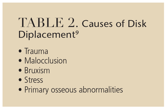
Disk displacement or internal derangement is when the articular disk is out of position. There are many types of displacement and various causes of these derangements (Table 2), which make diagnosis and treatment challenging.9 Advances in diagnostic technologies, such as cone beam computed tomography and magnetic resonance imaging, have helped to make diagnosis more accurate.1
PARAFUNCTIONAL HABITS
Nonfunctional mandibular movements — such as bruxism, nail biting, finger/thumb sucking, speech alteration, mouth breathing, pacifier use, prolonged gum chewing and atypical swallowing — are associated with a variety of jaw muscle symptoms and disk derangements.10 Chronic parafunctional clenching is suspected in chronic and acute TMDs. These habits are typically more common among women than men, and may often increase in frequency and intensity when patients experience stress.11
key takeaways
- The masticatory system consists of the structures required for adequate chewing, including the jaw and its related muscles, teeth, temporomandibular joints, tongue, lips, cheeks and mucous membranes.
- With a basic understanding of relevant concepts and principles relating to the masticatory system and tooth movement, clinicians can alert patients to possible problems and plan appropriate treatment.
- Evidence of these types of failures may include worn or fractured teeth, fractured restorations, and sore and/or enlarged muscles of mastication.
- A patient’s maximum opening of the oral cavity is a good indicator of potential temporomandibular joint issues. If a patient has limited opening, there may be something out of balance with the masticatory system.
- Palpating the muscles of mastication during routine oral cancer screening is a simple strategy to check for masticatory system problems.
- Because occlusal patterns and loads on natural teeth can vary over time as they shift, possibly affecting the placement of implants, patients with a combination of natural teeth and dental implants should have their occlusion periodically examined.
- By noting occlusal problems early, dental professionals can help patients avoid costly rehabilitation in the future.
ASSESSMENT
Treatments for TMD- or occlusion-related problems can range from a “watch and wait” approach to minor occlusal adjustments or the fabrication of occlusal guards or splints. Clinicians can help patients understand that something is out of balance with the masticatory system, and that problems may occur if left untreated. Evidence of these types of failures easily noticed during routine dental appointments may include worn or fractured teeth, fractured restorations, and sore and/or enlarged muscles of mastication.
Aspects of occlusal evaluation are simple to evaluate during recare appointments. A patient’s maximum opening is a good indicator of potential TMJ issues. Normal maximum opening is typically 40 mm to 50 mm, or about the width of four fingers. Most people can move their jaw to each side about 7 mm to 15 mm, and can protrude their jaw 10 mm to 15 mm.1 If a patient has limited opening, there may be something out of balance with the masticatory system.
Palpating the muscles of mastication during routine oral cancer screening is a simple strategy to check for masticatory system problems. Palpating the temporal, occipital, masseter, sternocleidomastoid, digastrics, trapezius, and pterygoid muscles and noting tenderness can indicate occlusal disharmonies. Again, educating the patient about the possible link between tenderness of these muscles and occlusal issues will encourage them to seek further diagnosis.
CONSIDERATIONS FOR DENTAL IMPLANTS![]()
Special attention should be given to the occlusal assessment of teeth restored with dental implants. With patients who have a combination of both natural teeth and dental implants, care should be taken to ensure that the dental implants are not overloaded. Teeth are supported and attached to the alveolar bone by the periodontal ligament. This soft tissue attachment to the hard, boney surface allows for a small amount of tooth movement. Movement up to 50 μm in any direction is considered normal in a healthy periodontium. Dental implants, on the other hand, have no soft tissue interface and are integrated directly with the alveolar bone. No movement with crowns supported by dental implants is considered normal. This difference in the dynamic interface of teeth and implants to alveolar bone means that the occlusal load will vary.
With patients who only have natural dentition, the occlusal contacts should be spread evenly, both in spacing and force, along all of the posterior teeth when the patient taps his or her teeth together straight up and down. Anterior teeth may or may not come in contact during this type of movement. If the patient has canine-protected occlusion, there should be no contact of posterior teeth during excursive or grinding movements.
This assessment can be accomplished by using articulating paper and having the patient first bite lightly, straight up and down. After marking the teeth during this movement, the dental professional should observe even-sized marks on the natural teeth and no marks on the crowns supported by implants. Next, have the patient strongly clench and grind his or her teeth side to side while the articulating paper is interposed. This time, the marks on the implant crowns located in the central grooves of the occlusal tables of posterior teeth and on the tips of the functional cusps should be noted. There should be no marks on cuspal inclines of any posterior teeth. If the dentist notices any inappropriate marks, appropriate occlusal adjustments can be made.12
During excursive movements, there should be no contact along cuspal inclines of crowns supported by dental implants. When the patient clenches tightly and grinds side to side, the periodontal ligament is compressed and the tooth actually depresses slightly in the socket. This is the reason for the lighter occlusal pattern that should be placed on dental implant crowns.
Because occlusal patterns and loads on natural teeth can vary over time as they shift and move, possibly affecting the placement of implants, patients with a combination of natural teeth and dental implants should have their occlusion periodically examined.
CONCLUSION
Patient education and a few simple checks can avert many problems associated with occlusal issues. By noting these types of problems early and encouraging patients to address them and comply with treatment, dental professionals can help patients avoid costly rehabilitation in the future.
References
- Dawson PE. Functional Occlusion: From TMJ to Smile Design. Philadelphia: Mosby; 2006:6,59,229,271.
- Goldstein BH. Temporomandibular disorders: a review of current understanding. Oral Surg Oral Med Oral Pathol Oral Radiol Endod. 1999;88:379–385.
- McNeil C. Management of temporomandibular disorders: concepts and controversies. J Prosthet Dent. 1997;77:510–522.
- Johansson A, Unell L, Carlsson GE, Söderfeldt B, Halling A. Gender difference in symptoms related to temporomandibular disorders in a population of 50-year-old subjects. J Orofac Pain. 2003;17:29–35.
- Macfarlane TV, Blinkhorn AS, Davies RM, Kincey J, Worthington HV. Orofacial pain in the community: prevalence and associated impact. Community Dent Oral Epidemiol. 2002;30:52–60.
- Pow EH, Leung KC, McMillan AS. Prevalence of symptoms associated with temporomandibular disorders in Hong Kong Chinese. J Orofac Pain. 2002;15:228–234.
- Goulet JP, Lavigne GJ, Lund JP. Jaw pain prevalence among French-speaking Canadians in Quebec and related symptoms of temporomandibular disorders. J Dent Res. 1995;74:1738–1744.
- National Institute of Dental and Craniofacial Research. Temporomandibular Joint and Muscle Disorders. Available at: www.nidcr.nih.gov/OralHealth/Topics/TMJ/. Accessed November 9, 2015.
- Berquist T. MRI of the Musculoskeletal System. 4th ed. Baltimore: Lippincott Williams & Wilkins, 2000.
- Castelo PM, Gavião MB, Pereira LJ, Bonjardim LR. Relationship between oral parafunctional/nutritive sucking habits and temporomandibular joint dysfunction in primary dentition. Int J Paediatr Dent. 2005;15:29–36.
- Bhat S. The etiology of temporomandibular disorders: the journey so far. International Dentistry. 2010;4:88–92.
- Misch CE. Contemporary Implant Dentistry. 2nd ed. St. Louis: Mosby Inc; 1999:151–162.
