

Conservative Management of a Single Dark Tooth
When formulating a treatment plan, clinicians must understand the etiology of discolored teeth and carefully weigh all clinical options.
When formulating a treatment plan, clinicians must understand the etiology of discolored teeth and carefully weigh all clinical options
Alterations in tooth color in the esthetic zone can prove challenging for restorative dentists who seek to provide conservative treatment. Achieving an “ideal” result can be difficult, and selecting a conservative approach requires thorough knowledge of minimally invasive procedures. In 1951, Markley1 stated that “loss of even part of a human tooth should be considered a serious injury, and that dentistry’s goal should be to preserve healthy natural tooth structure.”
As in all aspects of dentistry, diagnosis plays an important role in choosing the best treatment option. In these cases, it is necessary to understand the etiology of tooth discoloration. Historically, discoloration has been classified according to the location of the stain, which may be either intrinsic or extrinsic.2 Intrinsic discoloration occurs when the structural composition or thickness of the dental hard tissues are altered; extrinsic discoloration is caused by organic stains on the enamel surface.2 Additionally, any histological changes in enamel, dentin or coronal pulp structures can affect the light-transmitting properties of the tooth,3 modifying its natural appearance. Common local causes of tooth discoloration include pulpal necrosis, intrapulpal hemorrhage, pulp tissue remnants after endodontic therapy, endodontic materials, coronal restorative materials, root resorption and aging.4
Another common cause of tooth discoloration is pulp canal obliteration (PCO). Classified as either partial or total,5 this is normally caused by a traumatic injury.6 Severe trauma may cause pulpal necrosis, while moderate injury may cause PCO.7 Pulpal necrosis as a consequence of PCO has been found to be rare. Andreasen et al7 reported this occurrence in less than 1% of cases, while Oginni et al8 found this occurrence to be higher in teeth with total PCO (43%) compared to partial PCO (14%). Some studies have identified reduced transparency or yellowish discoloration of the tooth crown, regardless of the degree of obliteration.4,6 In the clinical experience of the authors, the degree of obliteration of the pulp chamber has a direct correlation to the intensity of color change, which can range from mild yellowish to dark yellow/brown.
The purpose of this clinical report is to present a simple and conservative treatment approach for the esthetic management of dark teeth with different etiologies in the same patient.
CLINICAL REPORT
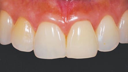
A 40-year-old Hispanic female with unremarkable medical history presented with a concern that her left maxillary central incisor (#9) and right maxillary lateral incisor (#7) were discolored (Figure 1 and Figure 2). Her dental history revealed previous orthodontic treatment in her teenage years, and she attributed her tooth discoloration to the orthodontic treatment. A clinical examination of the area of concern revealed a composite restoration on the lingual fossa (access for the canal) and Class III composite restorations on the mesiolingual and distolingual surfaces of the central incisor (#9), while the lateral incisor (#7) was intact. The patient was caries-free and maintained good oral hygiene and regular prophylaxes. Periapical radiographs revealed root canal treatment, with a 10-plus-year history on the central incisor (#9), and PCO on the lateral incisor (#7). The patient had no recollection of trauma to this area. In addition, no periapical lesions or widening of the periodontal ligament space were found (Figures 3A and 3B).

The patient had an endodontic consultation, which included an intraoral exam, shifted periapical radiographs, and a cone beam computed tomographic image to assess the presence and type of calcification. Electrical pulp testing of the right maxillary lateral incisor (#7) was within normal limits, but the tooth exhibited a delayed response to cold. A diagnosis of partial PCO with a normal periapex was established, with no treatment indicated at this time. Due to the patient’s desire to conserve tooth structure, a minimally invasive treatment option was presented that consisted of nightguard bleaching.

A horseshoe-shaped stone cast was made from an irreversible hydrocolloid impression of the patient’s maxillary arch to facilitate fabrication of a vacuum-formed thermoplastic tray for use as the bleaching carrier. The bleaching process was divided into two phases. The first phase included use of a modified single-tooth tray so that both dark teeth were isolated from the rest of the arch and bleached at the same time until they matched the adjacent teeth. The second phase included use of a maxillary full-arch tray to stabilize and achieve uniformity in the shades across the arch.
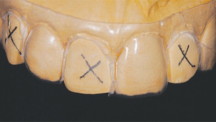
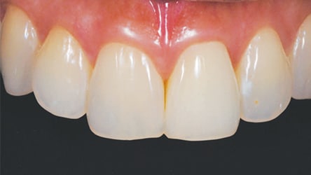
The design for the single-tooth tray was nonscalloped, nonreservoir, with the teeth molds on either side of the discolored teeth cut out (Figure 4). An “X” was placed on the stone cast on the right canine (#6), right central incisor (#8), and left lateral incisor (#10) to ensure the tray was cut out in those areas. The design for the full-arch tray was also nonscalloped, nonreservoir and covered all teeth in the patient’s maxillary arch. The patient was given 10% carbamide peroxide bleaching gel and instructed to use the single-tooth tray, only applying bleaching gel to the right lateral incisor (#7) and left central incisor (#9), and to wear the tray overnight for two weeks. At the conclusion of phase one, the shades of the right lateral incisor (#7) and left central incisor (#9) matched the adjacent teeth (Figure 5). The patient was instructed to use the full-arch tray and apply the bleaching gel to all maxillary teeth, wearing the tray overnight for an additional two weeks. She reported no sensitivity or adverse side effects during the duration of the bleaching. At the conclusion of phase two, a uniform shade was achieved to the patient’s satisfaction (Figure 6 and Figure 7).

DISCUSSION
Discolored teeth are a common concern in general dentistry practice, and patients do not always know the causative factor. The individual in this clinical report claimed the causative factor of her discolored teeth was orthodontic treatment, yet few studies have investigated a possible correlation between orthodontic treatment and PCO. In these studies, no evidence has been found to suggest that normal orthodontic forces contribute to PCO — but excessive forces cannot be ruled out as a potential cause.9,10 In this clinical report, however, there is no radiographic evidence of blunting of the apical portion of the roots or other signs of excessive orthodontic forces. However, the presence of previous endodontic therapy on the left maxillary incisor (#9) — without obvious extensive decay/restoration — suggests the possibility of past trauma to the tooth. Root canal treatment of the right maxillary lateral incisor (#7) was not recommended because most of the literature does not support endodontic intervention unless periradicular pathosis is detected or the involved tooth becomes symptomatic.11

There are numerous treatment options for the esthetic improvement of discolored teeth, including procedures involving composite restorations, veneers, crowns, or internal bleaching in nonvital or endodontically treated teeth. However, restorative options that consist of unnecessary, excessive removal of tooth structure could be considered overtreatment. For example, if veneers had been the treatment plan of choice for this patient, an estimated 0.7 to 0.9 mm of facial reduction would have been required to provide enough material thickness to mask the discolored teeth. Additionally, in order for the results to be esthetically pleasing, it may have been necessary to place veneers on all teeth in the esthetic zone so the shades and appearance would be consistent. This patient was concerned about loss of tooth structure, so the most conservative treatment option — nightguard bleaching with 10% carbamide peroxide — was selected for both discolored teeth. Nightguard bleaching was also appealing due to its low cost, safety, effectiveness12,13 and simplicity. While internal bleaching can be used for lightening endodontically treated teeth, with the removal of the lingual access restoration, it presents the risk of loss of tooth structure. Furthermore, approximately 7% of internal bleaching cases will develop external resorption defects.14,15 Regardless of the bleaching technique used, a relapse is possible, and is best addressed by external bleaching in a single-tooth tray with 10% carbamide peroxide to rebleach the tooth until it matches the surrounding dentition.16
In summary, discolored teeth can present numerous clinical challenges, ranging from determining etiology to formulating an appropriate treatment plan. The patient in this report was offered a conservative resolution to her chief complaint through simple nightguard bleaching. The results were pleasing to the patient, and no tooth structure was sacrificed. In a time in which operative treatment seems to be the default for esthetic dentistry, it is important to remember that more conservative options are often available.
KEY TAKEAWAYS
- Histological changes in enamel, dentin or coronal pulp structures can affect a tooth’s light-transmitting properties.3
- Causes of tooth discoloration commonly include pulpal necrosis, intrapulpal hemorrhage, pulp tissue remnants after endodontic therapy, endodontic materials, coronal restorative materials, root resorption or aging.4
- Another common cause of tooth discoloration is partial or total pulp canal obliteration, which normally results from trauma.
- Clinicians must understand the etiology of discolored teeth and carefully weigh all clinical options — including nightguard bleaching — before initiating treatment.
REFERENCES
- Markley MR. Restorations of silver amalgam. J Am Dent Assoc. 1951;43:133–146.
- Watts A, Addy M. Tooth discolouration and staining: a review of the literature. Br Dent J. 2001;190:309–316.
- Schaefer O, Watts DC, Sigusch BW, Kuepper H, Guentsch A. Marginal and internal fit of pressed lithium disilicate partial crowns in vitro: a three-dimensional analysis of accuracy and reproducibility. Dent Mater. 2012;28:320–326.
- Plotino G, Buono L, Grande NM, Pameijer CH, Somma F. Nonvital tooth bleaching: a review of the literature and clinical procedures. J Endod. 2008;34:394–407.
- Andreasen JO. Luxation of permanent teeth due to trauma. A clinical and radiographic follow-up study of 189 injured teeth. Scand J Dent Res. 1970;78:273–286.
- Jacobsen I, Kerekes K. Long-term prognosis of traumatized permanent anterior teeth showing calcifying processes in the pulp cavity. Scand J Dent Res. 1977;85:588–598.
- Andreasen FM, Zhijie Y, Thomsen BL, Andersen PK. Occurrence of pulp canal obliteration after luxation injuries in the permanent dentition. Endod Dent Traumatol. 1987;3:103–115.
- Oginni AO, Adekoya-Sofowora CA, Kolawole KA. Evaluation of radiographs, clinical signs and symptoms associated with pulp canal obliteration: an aid to treatment decision. Dent Traumatol. 2009;25:620–625.
- Delivanis HP, Sauer GJ. Incidence of canal calcification in the orthodontic patient. Am J Orthod. 1982;82:58–61.
- Popp TW, Artun J, Linge L. Pulpal response to orthodontic tooth movement in adolescents: a radiographic study. Am J Orthod Dentofacial Orthop. 1992;101:228–233.
- Amir FA, Gutmann JL, Witherspoon DE. Calcific metamorphosis: a challenge in endodontic diagnosis and treatment. Quintessence Int. 2001;32:447–455.
- Haywood VB. Considerations for vital nightguard tooth belaching with 10% carbamide peroxide after nearly 20 years of proven use. Inside Dentistry. 2006;2:2–5.
- Ritter AV, Leonard RH Jr, St Georges AJ, Caplan DJ, Haywood VB. Safety and stability of nightguard vital bleaching: 9 to 12 years post-treatment. J Esthet Restor Dent. 2002;14:275–285.
- Schwartz RS, Summitt JB, Robbins JW. Fundamentals of Operative Dentistry: A Contemporary Approach. Chicago, Ill: Quintessence Publishing; 1996.
- Todd M, Brackett W, Romero M. Correction of a single discolored anterior tooth due to internal resorption: a clinical report. Compend Contin Educ Dent. 2017;38:e13–e16.
- Haywood VB, DiAngelis AJ. Bleaching the single dark tooth. Inside Dentistry. 2010;6:42–52.
FEATURED IMAGE BY ARTPIPI/ISTOCK/GETTY IMAGES PLUS
The authors have no commercial conflicts of interest to disclose.
From Decisions in Dentistry. December 2017;3(12):22—25.
