
Clinical Considerations for Implant Maintenance
An exploration of peri-implant disease and therapies designed to help ensure successful implant outcomes.
An exploration of peri-implant disease and therapies designed to help ensure successful implant outcomes
The discovery of osseointegration revolutionized dentistry. The ability to replace teeth in a stable, predictable way has changed the way endentulous patients are treated. Unfortunately, even under optimum circumstances, implant failures occur. While the specific etiology of failures has not been completely elucidated, it has been shown that biofilms and micromotion (associated with normal mastication or bruxing) can have negative effects on the longevity of these devices.1 If one accepts these two factors — or a combination thereof — as being among the primary etiologic reasons for implant loss, then it behooves clinicians to evaluate the health of the peri-implant tissues, as well as the occlusion, on implants. This requires proper planning, placement and restoration of these fixtures. Once the final prosthetics are in place, maintenance is appropriate, because, to some degree, biofilm and occlusal forces can be controlled.1
Proper planning includes selecting an appropriate patient, and choosing the appropriate diagnostic tools, implant software and implant system. Adequate training and expertise have been demonstrated to influence implant success.2 Optimum selection of restorative design, materials and loading protocols must also be considered.3–5 In spite of strict application of these criteria, peri-implant disease can occur. It has been estimated that five to 10 years after placement, 10% of implants in 20% of patients will have an inflammatory process around the devices.6 One recent study indicated that, at nine years, as many as 45% of implant sites may exhibit signs of inflammation.7 These inflammatory conditions have been classified as peri-implant mucositis and peri-implantitis.
Peri-implant mucositis is inflammation of the peri-implant soft tissues without progressive bone loss. Peri-implantitis is inflammation characterized by increased probing depths with bleeding and/or suppuration associated with progressive bone loss. It is correct to assume that oral biofilm responsible for the etiopathogenesis of periodontal disease that colonizes all hard surfaces in the oral cavity will also colonize dental implant surfaces and their restorative components. Biofilm has been shown to produce peri-implant mucositis, which, if left untreated, can progress to peri-implantitis.8 Dental biofilm can affect osseointegration not just by inducing an inflammatory response, but also by altering the implant surface itself. While multiple materials are used for the fabrication of implants, titanium is, by far, the most common implant material (and, for the purposes of this paper, it is assumed that the implants in question are made of titanium). It is therefore important to evaluate the behavior of these materials when exposed to the oral cavity.
TITANIUM OXIDE LAYER
Titanium osseointegrates due to its passivation properties by which a titanium oxide (TiO) layer forms on the surface of the implant. If the titanium surface is covered with microbial biofilm byproducts, the availability of oxygen decreases. If this happens in combination with abnormal forces, the oxide layer is continuously removed, and if oxygen is not present in abundance, repassivation reactions are not favored.9,10
This altered surface cannot be reconstituted in vivo using current approaches. The titanium oxide layer can be removed or destroyed in the early stages of therapy. The action of microorganisms common to the oral cavity, including Streptococcus mutans, Candida albicans and Porphyromonas gingivalis, can produce acids capable of eroding the TiO layer.11 This finding alone would justify routine maintenance for patients with dental implants. The challenge is how to treat the contaminated implant surface. Oral hygiene instructions and appropriate plaque control can help, and should be discussed with patients to help them effectively control biofilm between maintenance visits.
OCCLUSAL FORCES

Abnormal forces can negatively affect the implant surface. It is well known that excess macromotion (trauma) can fracture implants (Figure 1). Recent studies have raised concerns, however, about the micromotion caused by routine mastication and parafunctional habits, including clenching and bruxing. Research has shown that micromotion alone can lead to deterioration of the titanium oxide layer.12
It is therefore important to control the occlusal forces on implants, as these devices function best under vertical loads. Obtaining these vectors requires precise planning, placement and restoration. Optimal emergence of the long axis of the implant abutment in the center of the fixed partial denture in the posterior is recommended. In general, posterior implants function best with light contact in centric relation, and no contact in lateral excursive movements. In a patient with both implants and teeth, lateral forces should be placed on teeth where possible, thus reducing the load on implants. Relieving pressure from parafunctional habits is best accomplished by fabricating a maxillary, full-coverage, hard-acrylic occlusal guard. For the average patient, this device will allow freedom from centric relation to centric occlusion, with lateral and protrusive excursions picked up by the mandibular canines. At maintenance visits, occlusal guards should be evaluated for proper contact and inappropriate wear on occlusal surfaces.
When biofilm and micromotion are combined, the alteration of the TiO layer previously described has been shown to occur at a more rapid rate.11 The combination leads to a phenomenon called tribocorrosion. This is defined as a surface transformation resulting from the interactions of mechanical loading and chemical reactions. This corrosion has been associated with particles of titanium being deposited into soft tissues.13 These titanium ions have also been associated with the initiation of inflammatory processes in soft tissues in orthopedics.14 At present, the role that particles of titanium — and titanium ions in soft tissue — play in the genesis and continuation of peri-implant disease is unclear. Because the in vitro studies previously cited have demonstrated that a combination of biofilm and occlusal forces can release this material, it seems reasonable these parameters should be checked on a routine basis.
In most cases, the early stages of peri-implantitis are asymptomatic. Thus, periodic radiographic examinations should be performed to evaluate bone changes at the marginal level. Occasionally, excess cement on implants restored with cemented crowns can be detected on radiographs. This can only be assessed if the cement used is radiopaque, thick, and on the mesial or distal surface of the implant. Excess cement is associated with peri-implant disease;15 more specifically, small pieces of luting agents have been associated with inflammation, and if excess cement is not removed, it can lead to implant loss. In most cases, this inflammation doesn’t appear until months or, more commonly, several years after cementation.15
Key Takeaways
- It has been estimated that five to 10 years after placement, 10% of implants in 20% of patients will have inflammation.6 One recent study indicated that, at nine years, as many as 45% of implants may exhibit signs of inflammation.7
- These inflammatory conditions are classified as either periimplant mucositis or peri-implantitis.
- Dental biofilm can affect osseointegration not just by inducing an inflammatory response, but also by altering the implant surface itself.
- Recent studies have also raised concerns that micromotion — caused by routine mastication and parafunctional habits, including clenching and bruxing — can lead to deterioration of the implant’s titanium oxide layer.12
- Excess cement can contribute to peri-implant disease.15 Small pieces of luting agents have been associated with inflammation, and if excess cement is not removed, it can lead to implant loss.
- There is no consensus about the frequency of implant maintenance visits.21 These visits can usually be set using two parameters: the health of the patient’s dentition, and health of the peri-implant tissues.
BLEEDING ON PROBING
Bleeding on probing (BOP) is an early sign of cement associated with peri-implant disease. That said, BOP is not unusual around dental implants, and is often simply associated with poor oral hygiene. When BOP around an implant is detected, it is suggested that the sulcus be gently cleaned, the patient rinse twice daily with chlorhexidine for 30 days, and that clinicians reinforce the patient’s personal oral hygiene instructions. The patient should be reevaluated after 30 days. If bleeding is still evident after probing around a cemented restoration, excess cement is likely present. In most cases, complete removal of subgingival remnants without direct visibility is difficult. Frequently, periodontal endoscopy is used to identify cement deposits. The cement should be removed as completely and expeditiously as possible. If BOP persists after nonsurgical attempts, surgical intervention may be warranted. Only with routine evaluations can early signs of inflammation be found and treated, thus reducing the probability of implant loss. This again argues for appropriate maintenance for these patients.
ROUTINE IMPLANT MAINTENANCE
Maintenance regimens for patients with implants closely resemble those indicated for dentate individuals. A review of the patient’s systemic health, medications, hospitalizations and other health care data since the last visit is appropriate. Tobacco use should be discouraged because it has been associated with an increased risk of peri-implantitis and poor prognosis.16 This is followed by a routine extraoral examination for any abnormalities, including an evaluation of peri-implant soft tissues. A routine oral cancer screening should be performed. There are case reports in the literature of neoplasia or metastasis found in close proximity to implants, mimicking peri-implant inflammatory conditions.17–20

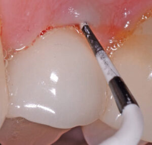
Oral hygiene should be evaluated, and the patient should receive specific instructions about adjunct devices or techniques that would better help to remove plaque and food debris to prevent inflammation.21,22 Probing depths similar to those taken around teeth (six per fixture) are recorded. This can be performed with a plastic probe (Figure 2), however, due to the various configurations of the restorative components for fully or partially edentulous patients, probing around implants can by challenging. Gentle probing with a metallic probe may be performed. When appropriate, right-angle radiographs are exposed. These are compared to those taken immediately after implant placement and after final restoration of the fixture. If a comparison of bone levels reveals any marginal bone loss, the etiology of this process should be diagnosed and treated as soon as possible. Following a routine examination of the remaining dentition, the patient’s oral hygiene regimen should be evaluated and modified, as appropriate. Occlusion should also be evaluated. The goal is to identify and eliminate, if possible, horizontal forces on implants. In addition, the patient’s occlusal guard can be evaluated for proper fit and to eliminate any inappropriate contacts.

Cleaning around implants without altering the implant surface can be difficult. For routine cleaning around healthy implants, recent research suggests that the use of metallic curets, as well as sonic or ultrasonic instruments, should be avoided because these devices may result in titanium particles being deposited into the surrounding soft tissues.13 According to animal studies and human studies, pathogenic bacteria and their byproducts can be removed by rubbing the implant surface using sterile saline or chlorhexidine on cotton gauze (Figure 3).23,24
The surface of the implant should be gently rubbed for no more than five minutes to avoid removing excess amounts of titanium particles and ions.25 Nonmetallic curets can be used for biofilm removal (Figure 4). After removing bacterial plaque and calculus from the abutment or implant, the surface can be polished using rubber cups to restore smoothness and prevent additional plaque accumulation.23 Removing excess cement is best accomplished when the original restoration is placed. After that, the complete removal of residual deposits becomes difficult. In these cases, the use of a periodontal endoscope, dental videoscope, or open flap surgery is frequently needed. The goal is removal of excess cement on the implant, restoration and from the soft tissues.
MAINTENANCE FREQUENCY
At present, there is no consensus about the frequency of implant maintenance visits.21 These visits can usually be set using two parameters: the health of the patient’s dentition, and health of the peri-implant tissues. For patients who have no signs of inflammation around either implants or teeth, an annual evaluation is appropriate, whereas individuals with gingivitis, but with healthy peri-implant tissues, can be seen twice a year. Patients with complex or incorrect prosthetic design may need to be seen more often to assist these individuals with adequate debridement.26 Patients presenting with periodontitis, but no peri-implant disease, should be seen every two to three months (keeping in mind that previous history of periodontitis is considered the main risk factor for peri-implantitis).27 Patients with peri-implantitis should be appointed to find the genesis of the disease and provide appropriate treatment.
At present, there is no consensus about the frequency of implant maintenance visits
While the profession continues to learn more about why dental implants fail, current data strongly suggest that routine maintenance for these devices is appropriate.
References
- Wilson TG Jr, Valderrama P, Rodrigues DBC. The case for routine maintenance of dental implants. J Periodontol. 2014;85:657–660.
- Da Silva, JD, Kazimiroff J, Papas A, et al. Outcomes of implants and restorations placed in general dental practices. J Am Dent Assoc. 2014;145:704–713.
- Sanz-Sánchez I, Sanz-Martín I, Figuero E, Sanz M. Clinical efficacy of immediate implant loading protocols compared to conventional loading depending on the type of the restoration: a systematic review. Clin Oral Implants Res. 2015;26:964–982.
- Zygogiannis K, Wismeijer D, Aartman IH, Osman RB. A systematic review on immediate loading of implants used to support overdentures opposed by conventional prostheses: factors that might influence clinical outcomes. Int J Oral Maxillofac Implants. 2016;31:63–72.
- Muddugangadhar BC, Armarnath GS, Sonika R, Chheda PS, Garg A. Meta-analysis of failure and survival rate of implant-supported single crowns, fixed partial denture, and implant tooth-supported prostheses. J Int Oral Health. 2015;7:11–17.
- Mombelli A, Müller N, Cionca N. The epidemiology of peri-implantitis. Clin Oral Implants Res. 2012;23:67–76.
- Derks J, Schaller D, Håkansson J, Wennstrom JL, Tomasi C, Berglundh T. Effectiveness of implant therapy analyzed in a Swedish population: Prevalence of peri-implantitis. J Dent Res. 2016;95:43–49.
- Pontoriero R, Tonelli MP, Carnevale G, Mombelli A, Nyman SR, Lang NP. Experimentally induced peri-implant mucositis: a clinical study in humans. Clin Oral Implants Res. 1994;4:254–259.
- Rodrigues DC, Urban RM, Jacobs JJ, Gilbert JL. In vivo severe corrosion and hydrogen embrittlement of retrieved modular body titanium alloy hip-implants. J Biomed Mater Res B Appl Biomater. 2009;88:206–219.
- Sridhar S, Abidi Z, Wilson TG Jr, et al. In vitro evaluation of the effects of multiple oral factors on dental implant surfaces. J Oral Implantol. 2016;42:248–257.
- Rodrigues DC, Sridhar S, Gindri I, et al. Spectroscopic and microscopic investigation of the effects of bacteria on dental implant surfaces. RSC Adv. 2016;6:48283–48293.
- Sridhar S, Wilson TG Jr, Palmer KL, et al. In vitro investigation of the effect of oral bacteria in the surface oxidation of dental implants. Clin Imp Dent Rel Res. 2015;17: e562–e575.
- Wilson TG Jr, Valderrama P, Burbano M, et al. Foreign bodies associated with peri-implantitis human biopsies. J Periodontol. 2015;86:9–15.
- Pettersson M, Kelk P, Belibasakis GN, Bylund D, Molin Thorén M, Johansson A. Titanium ions from particles that activate and execute interleukin-1 release from lipopolysaccharide-primed macrophages. J Periodont Res. March 14, 2016. Epub ahead of print.
- Wilson TG Jr. The positive relationship between excess cement and peri-implant disease: A prospective clinical endoscopic study. J Periodontol. 2009;80:1388–1392.
- de Waal YC, Raghoebar GM, Meijer HJ, Winkel EG, van Winkelhoff AJ. Prognostic indicators for surgical peri-implantitis treatment. Clin Oral Implants Res. March 29, 2015. Epub ahead of print.
- Pfammatter C, Lindenmüller IH, Lugli A, Filippi A, Kühl S. Metastases and primary tumors around dental implants: a literature review and case report of peri-implant pulmonary metastasis. Quintessence Int. 2012;43:563–570.
- Raiser V, Abu-El Naaj I, Shlomi B, Fliss DM, Kaplan I. Primary oral malignancy imitating peri-implantis. J Oral Maxillofac Surg. 2016;74:1383–1390.
- Jané-Salas E, López-López J, Roselló-Llabrés X, Rodríguez-Argueta OF, Chimenos-Küstner E. Relationship between oral cancer and implants: clinical cases and systemic literature review. Med Oral Patol Oral Cir Bucal. 2012;17:e23–e28.
- Moergel M, Karbach J, Kunkel M, Wagner W. Oral squamous cell carcinoma in the vicinity of dental implants. Clin Oral Investig. 2014;18:277–284.
- Armitage GC, Xenoudi P. Post-treatment supportive care for the natural dentition and dental implants. Periodontol 2000. 2016;71:164–184.
- Jepsen S, Berglundh T, Genco R, et al. Primary prevention of peri-implantitis: managing peri-implant mucositis. J Clin Periodontol. 2015;42:S152–S157.
- Valderrama P, Blansett JA, Gonzalez MG, Cantu MG, Wilson TG. Detoxification of implant surfaces affected by peri-implant disease: an overview of non-surgical methods. Open Dent J. 2014;8:77–84.
- Schwartz F, John G, Becker J. Reentry after combined surgical respective and regenerative therapy of advanced peri-implantitis: a retrospective analysis of five cases. Int J Periodontics Restorative Dent. 2015;35:647–653.
- Wheelis SE, Gindri IM, Valderrama P, Wilson TG Jr, Huang J, Rodrigues DC. Effects of decontamination solutions on the surface of titanium: investigation of surface morphology, composition, and roughness. Clin Oral Implants Res. 2016;27:329–340.
- Canullo L, Schlee M, Wagner W, Covani U; Montegrotto Group for the Study of Peri-Implant Disease. International brainstorming meeting on etiologic and risk factors of peri-implantitis, Montegrotto (Padua, Italy), August 2014. Int J Oral Maxillofac Implants. 2015;30:1093–1104.
- Eick S, Ramseier CA, Rothenberger K, Brägger U, Buser D, Salvi GE. Microbiota at teeth and implants in partially edentulous patients. A 10-year retrospective study. Clin Oral Implants Res. 2016;27:218–225.
From Decisions in Dentistry. August 2016;2(08):22,24,26–29.
